Medical Models
We Build Custom 3D medical models for clinicians, health systems and healthcare vendors.
Table of Contents
Medical Models + Technology
Even though 3D Printing has been available for 35 years in industrial applications, it has recently caught the attention of healthcare professionals in creating medical anatomical models, implants, and medical devices. Clinicians, health systems, and vendors are all trying to understand how to best utilize and implement this technology into their roadmaps.
It is often hard to visualize aspects of a tumor or damage to soft/hard tissue looking at a CAT Scan or MRI. A physical 3D Printed medical anatomical model tells the entire story. Now, with 3D printing and other modern technologies, models are created in less time, are more affordable, more accurate, and have a higher level of detail than ever before. These 3D models provide a hands-on perspective with high detail and full-color segmentation, and are ideal for use used in pre-surgery planning and patient education.
Large 3D product replicas and models of medical devices, inventions and products are also valuable tools for trade shows, funding, demonstrations or prototyping.
Gallery of 3D Medical Models
Features and Benefits
- A physical model provides a visual perspective that you can‘t achieve with 2D sliced MRI/CT models.
- Models can be full-color, monochrome, transparent, pliable, and highly detailed. Strikingly high-resolution models with multiple options of finishes to emulate the desired outcome of the model such as dry-look or wet-look models.
- CT‘s and MRI‘s can be combined to do tractography studies.
- Models can be used for pre-surgery planning and practice, patient communication and education, prosthesis testing.
- Medical models are often used as courtroom exhibits for forensic visualization presentations to juries.
- Provides ability to interact in a learning environment with real case studies—demonstrating a prognosis and then comparing with the outcome is invaluable.
- Personalized anatomical models can be created fast with next-day service very economically.
- Tumor resection models can be used to clearly highlight the tumor and the surrounding tissue.
- Orthopedic models can be built from materials that are bone-like and can be used for pre-surgery measuring and medical device adjustments.
- Vascular models can be printed in full-color or transparent materials to identify abnormalities in the organ, tumors, blood flow, sliced chambers, valves, muscle tissue, calcified tissue and many other uses.
- Models can be used to combined both soft-tissue and hard-tissue.
- Medical models can be designed and built for viewing with sliced, hinged, magnetized, flexible, pull-aparts, and cutaway features.
Hinge and Slice Models
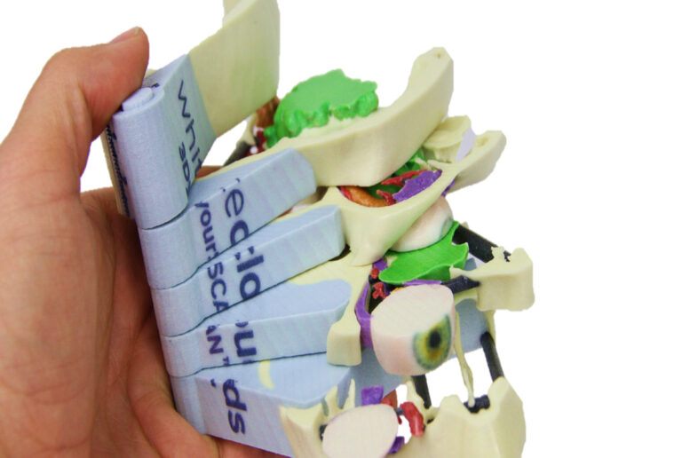
WhiteClouds has a patent-pending “Hinge and Slice” technology that allows for examining specific areas of interest in more detail by evaluating various slices of the model. These slices can be designed in varying thicknesses and view planes giving multiple views.
Steps in Creating the Medical Model
- Obtaining the DICOM file is the first step. These 2D images (slices) within DICOM files are produced from CT scans (Computed Tomography) or MRI scans (Magnetic Resonance Imaging).
- Since medical imaging techniques show the entire body, including the surrounding tissues, the 2D data set must be Segmented. Segmentation removes any undesired data within the region of interest so that only the relevant portions remain. There are several ways to accomplish this, but all typically require specialized software and trained personnel to perform an accurate segmentation.
- The outcome from the Segmentation step is a 3D model. A commonly overlooked step includes an engineering and functionality design to ensure the resulting 3D Print will have the proper support, pegging, slicing, presentation, color application, etc. This pre-production design of the 3D model is typically done by a 3D graphics designer.
- Once the 3D model has been completely designed, the file is output to an STL file (a typical 3D Printing file format). At this point, the file is 3D Printed on the appropriate printer and materials. There are many technologies that can 3D Print models in numerous materials allowing for full-color options, physical characteristics (from hard to pliable), and visual perspectives with transparent or hinge/slice models. Some of these technologies are FDM (Fused Deposition Modeling), CJP (Color Jet Printing), MJP (Multi-Jet Printing), PolyJet, SLA (Stereo Lithography Apparatus), SLS (Selective Laser Sintering) along with many others.
Medical Review Comments
“If you put a 3D model of a patient in the hands of a surgeon, they immediately realize the value. It helps in the education of patient and family, training of residents and fellows, and planning surgical approaches. It is the ultimate in patient-specific imaging.”
Dr. Edward P Quigley III MD Ph.D. Neuroradiology University of Utah
Dr. Jay Bishoff of Intermountain Healthcare, one of the nation’ leading urologic surgeons sees the technology making “good surgeons into great surgeons.” Continues Dr. Bishoff,
“These visual models revolutionize the way we perform surgery by providing insight that even the trained eye could not have seen before.” “Interactive 3D models are incredibly helpful to physicians, trainees, and patients for understanding anatomy,” said Dr. Justin Cramer of the University of Nebraska Medical Center. “Previously, full interaction with patient-specific models has been possible on expensive 3D software only available on special workstations. Our work has sought to make models readily available to anyone who wants them, and 3D printing is a large part of this effort. Tangible printed models facilitate interactive learning, and enable a more thorough understanding of the anatomy depicted.”
Dr. Jay Bishoff of Intermountain Healthcare
Pricing
Cost of medical models is based on the volume of material (size of the model), the time it takes to create the 3D printable file and other elements of the model. Each model is bid individually and the best way to determine cost is to email us, call us at 385-206-8700, or fill out the form below and let us bid on your project.
Get a Free Price Estimate for a Custom 3D Medical Model
Custom Fabrication Workflow
Technology and Materials
We can 3D print models in different materials including UV-cured resin. The type of model determines which material will produce the best results. We can help you choose the material that is best for your project.
3D printed medical models show incredible detail. The resolution of our printers is finer than a human hair.
Our patent-pending “Hinge and Slice” technology allows for examining specific areas of interest in more detail.
Common Questions & Answers
What file type is required to generate a 3D model?
Most medical model prints start with a DICOM set. However, the DICOM format is not a 3D format. The region of interested has to be segmented out of the DICOM slices and made into a 3D model. We can generate your model starting with your DICOM or we can start with a 3D file if you have already performed the segmentation.Do the printers print in color?
Yes. As requested, some models are printed in UV-cured resin materials with a transparent shell in the form of the internal organ. There are some color abilities with those materials but they are limited.Is color important?
Yes, for two reasons. First, there is often the need to visibly know the boundaries of certain regions. For example, the actual geometry of a tumor as compared to the tissue around it. Second, color adds realism to a medical model. This enhances communication with the patient, the family, and other medical professionals. Medical clinicians are accustomed to viewing a specific region of interested in a 3D viewer on their screen. In those viewers, a realistic color texture is applied for the above two reasons. Having the model in your hand look like what you are seeing on the screen is very important.What is the material used for medical models?
We print in UV-cured resins depending on the specific need.Can you make flexible, tissue-like medical models?
Yes. The colors will be limited but we can print in flexible materials.Can you make rigid, bone-like medical models?
Yes. Our material is treated in such a way that it feels somewhat like bone and can even be machined, sawed, drilled, etc. similar to bone using actual medical tools.What if I want to see inside the model?
It is common to want a medical model that has the form of the region of interest with the added requirement of being able see inside the model. For this purpose, we have a patent-pending method of creating slices of the model with an external hinge which allows the user to go to a specified depth to see the interaction of soft tissue, bone, vascular, etc.What is the turnaround time for a 3D-printed medical model?
We know the need for a medical model is often tied to an upcoming surgery. You typically have the model in your hands within 3 days.
Do you have a question we didn‘t answer? Don’t hesitate to contact us at 1-385-206-8700 or [email protected].
Worldwide Delivery
WhiteClouds has delivered models around the world.
History of Medical Models
Medical imaging is the technique and process of creating visual representations of the interior of a body for clinical analysis and medical intervention. Medical imaging has been used for hundreds (or thousands) of years. Many illuminated manuscripts and Arabic scholarly treatises of the medieval period contained illustrations representing various anatomical systems.
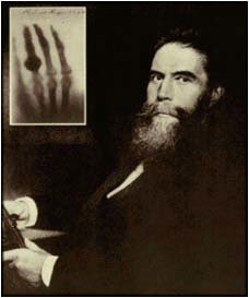
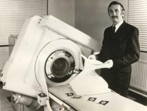
Modern medical imaging began with radiography after the discovery of x-rays in 1895 by Wilhelm Röntgen. In the 1970’s G.N. Hounsfield and A.M. Cormack were awarded the Nobel Prize in medicine for the invention of Computed Tomography (CT Scanning). In 1983, Michael W. Vannier, MD published a paper on his three-dimensional reconstruction of a human skull. As no computers existed yet to display three-dimensional CT scans, Vannier had to improvise: he used early CAD system to produce the 3D reconstruction. Modern volume rendering techniques have been developed to enable CT, MRI and ultrasound scanning software to produce 3D images for the physician.
Only in the last few years has 3D printing been applied to take these visualization techniques to the next level, allowing physicians the ability to visualize important structures in great detail, 3D visualization methods are a valuable resource for the diagnosis and surgical treatment of many pathologies.


