In the vast world of scientific exploration, there exists an extraordinary tool that uncovers a realm of hidden beauty, where the smallest of details transform into captivating works of art. Microscopy, the science of studying objects and structures too minuscule for the naked eye to behold, unveils a realm where beauty and scientific discovery converge in a symphony of colors, shapes, and intricate patterns. Join us on an exhilarating journey as we explore the stunning artistry found within the microscopic world.
Gecko Hand: The Marvel of Nature’s Engineering
Imagine a gecko adorning the walls, defying gravity with effortless grace. Such a spectacle is made possible by the extraordinary microscopic architecture of its feet. Grigorii Timin, an exceptionally talented scientist, in collaboration with the brilliant Dr. Michel Milinkovitch, delved into the mysteries of these captivating creatures. With the aid of microscopy, they uncovered the intricate details that allow geckos to walk on walls and ceilings effortlessly. Through the lens of their microscope, the gecko’s foot reveals a mesmerizing network of thousands of microscopic hairs, known as setae. These tiny structures employ van der Waals forces, enabling the gecko to stick to surfaces. Each seta branches into even smaller structures called spatulae, which create intimate molecular connections with the surface. Witnessing the intricate pattern of these hairs, one can’t help but marvel at the elegance of nature’s engineering.
Bee Antenna: A Symphony of Sensory Perception
The delicate beauty of a bee in flight is a sight to behold. ZEISS Microscopy, renowned pioneers in the field of microscopy, have captured the enchanting intricacy of a bee’s antenna. The bee’s antenna, beyond being a sensory marvel, showcases the remarkable power of microscopic exploration. Under the microscope, the bee antenna reveals a symphony of sensory perception. Thousands of tiny hairs called sensilla decorate the surface, each serving as a receptor for detecting scents, humidity, and even minute changes in temperature. As we immerse ourselves in the image, we witness the elegance and complexity of nature’s sensory architecture.
Acorn Barnacle: Nature’s Architectural Marvel
Beneath the ocean’s surface, a hidden world of architectural marvels awaits our exploration. Charles B. Krebs, a visionary in the realm of scientific imaging, has captured the awe-inspiring beauty of the acorn barnacle through his microscope lens. These tiny crustaceans adorn rocks, shells, and even the hulls of ships, creating captivating formations reminiscent of miniature towers. The microscope unveils a world of textured surfaces and intricate designs. Each barnacle, consisting of calcified plates, displays a stunning arrangement of overlapping structures. The captivating beauty lies not only in the overall form but also in the delicate textures and patterns revealed under the microscope’s watchful eye.
Southern Live Oak Leaf: A Tapestry of Life
Nature’s canvas, adorned with a rich tapestry of colors and shapes, has inspired artists for centuries. However, it is only through microscopy that we can truly appreciate the extraordinary details that make up this natural artwork. Jason Kirk, an accomplished photographer and microscopy enthusiast, has immortalized the mesmerizing beauty of a Southern Live Oak Leaf through his lens. Zooming in on the leaf’s intricate structure, we discover a breathtaking world of intricate patterns. The delicate veins intertwine, resembling a network of rivers and tributaries. Each cell, meticulously arranged and packed, contributes to the leaf’s strength and flexibility. The subtle shades of green dance in harmony, creating a captivating symphony of color.
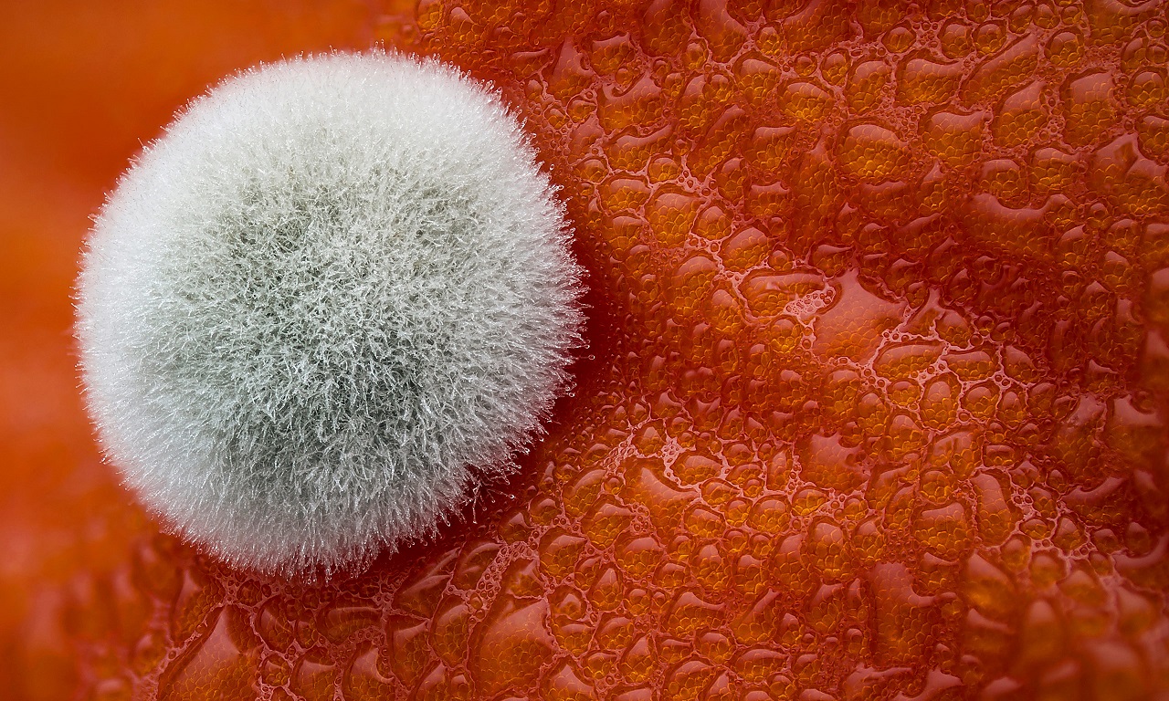
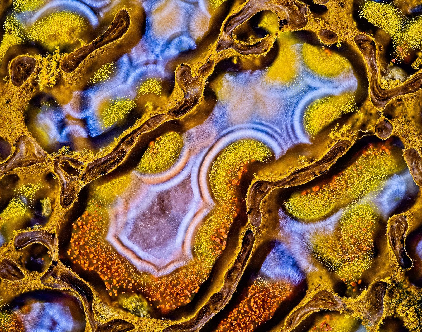
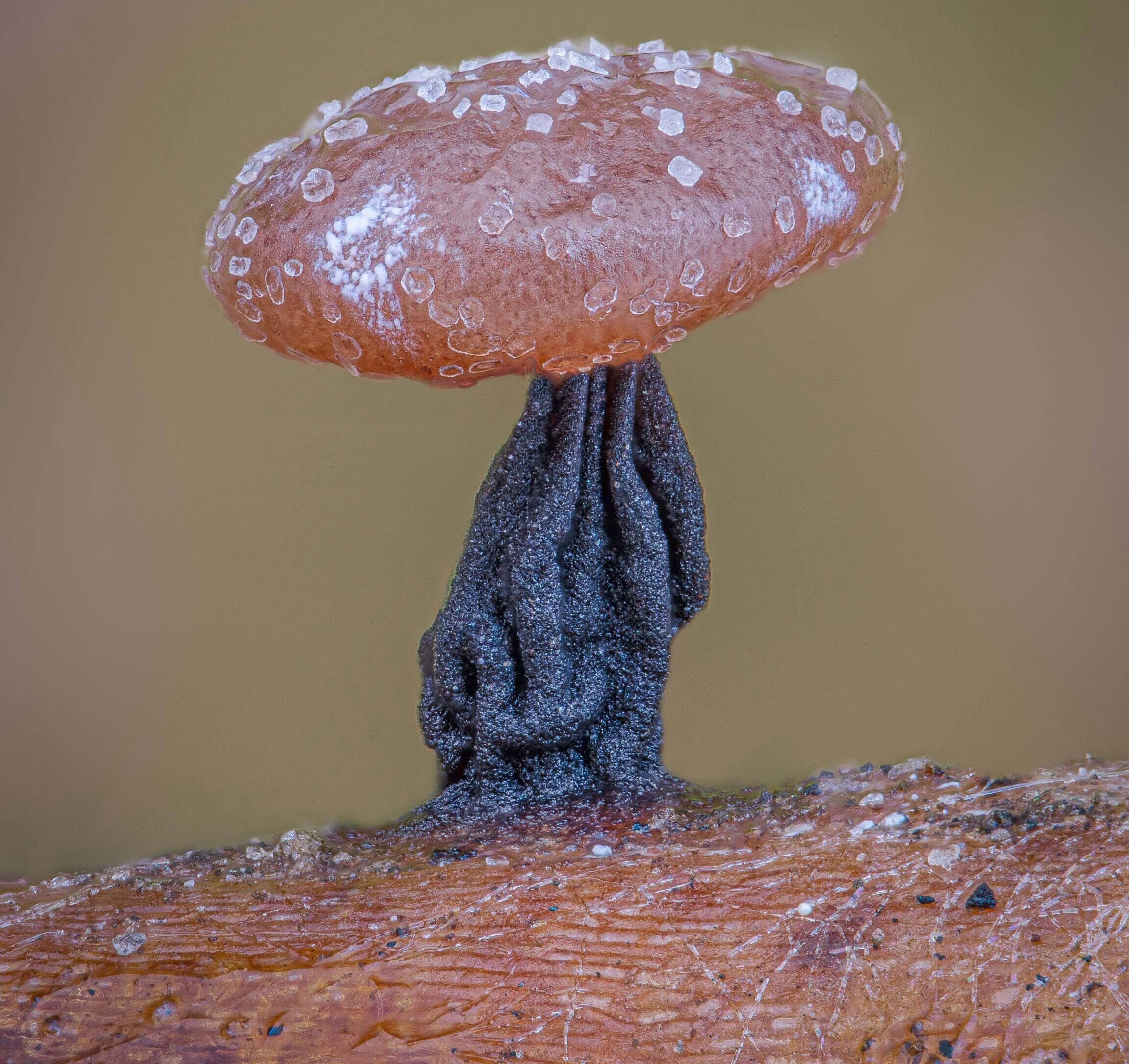
Mold on a Tomato: The Elegance in Decay
Even in decay, nature reveals its unique charm. Dean Lerman, a visionary explorer of the microscopic world, delved into the realm of decomposition and discovered the unexpected elegance in the mold that adorns a decaying tomato. Through his microscope, Lerman captured the intriguing beauty that emerges amidst the process of decay. At first glance, one might dismiss the sight of mold as unappealing or unsightly. However, under the microscope’s lens, a whole new world unfolds. The mold spores weave intricate networks of filaments, creating a mesmerizing display of interconnected patterns. Delicate hyphae extend like branches, colonizing the tomato’s surface in an elaborate dance of life and decay. As we gaze upon the mold-covered tomato, we witness a fascinating juxtaposition of colors and textures. Shades of blue, green, and white intertwine, forming a captivating mosaic. The mold, with its feathery structures and delicate formations, turns what some may perceive as rot into an unexpected work of art.
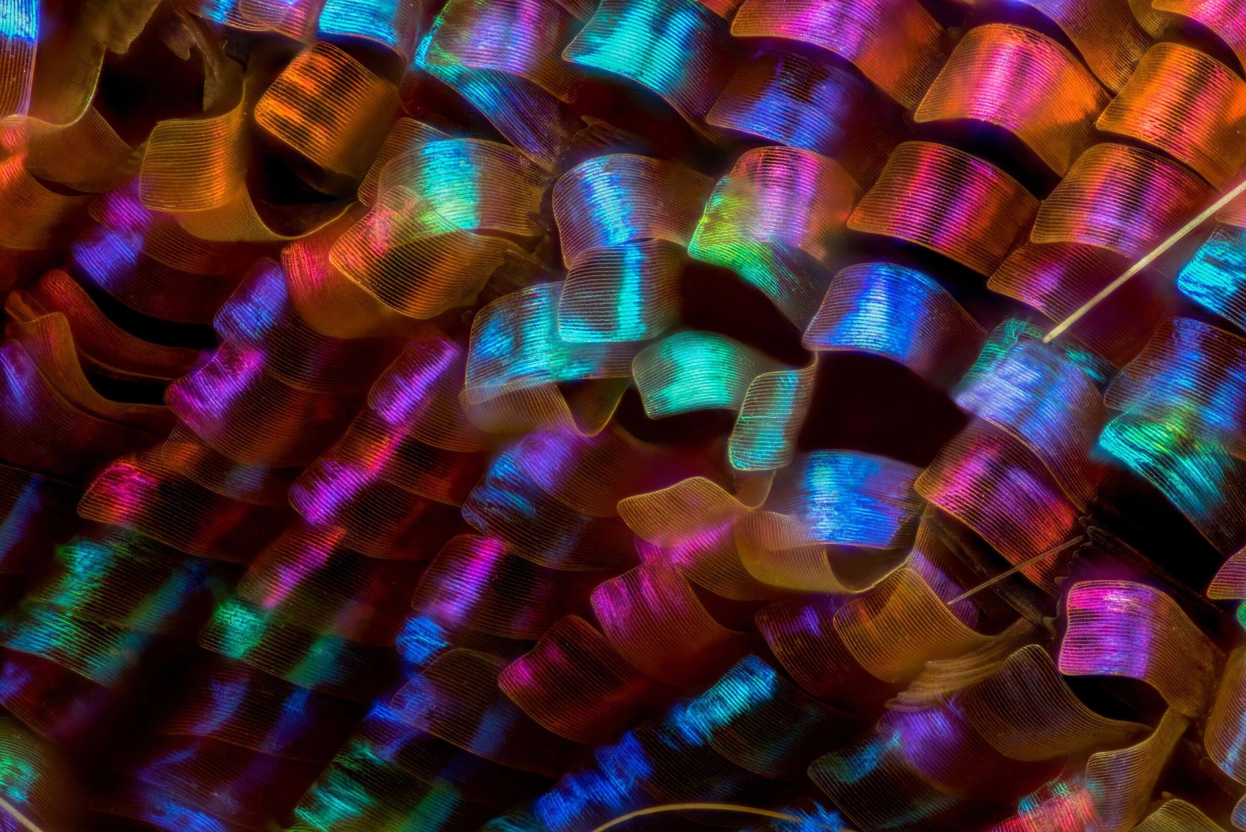
Cotton Fabric: The Woven Splendor
Dr. Felice Placenti, a visionary in the field of textile engineering, partnered with the power of microscopy to unlock the secrets of cotton fabric. Under the microscope’s lens, cotton fibers reveal an astonishingly intricate tapestry of interconnected threads. The fabric’s beauty lies not only in its softness and comfort but also in the complexity of its structure. As we zoom in, a world of intricate weaving patterns emerges. Each cotton fiber, composed of countless cellulose strands, intertwines with its neighbors, forming an exquisite network. The delicate textures, ranging from smooth to twisted, create a tactile symphony that showcases the mastery of human craftsmanship.
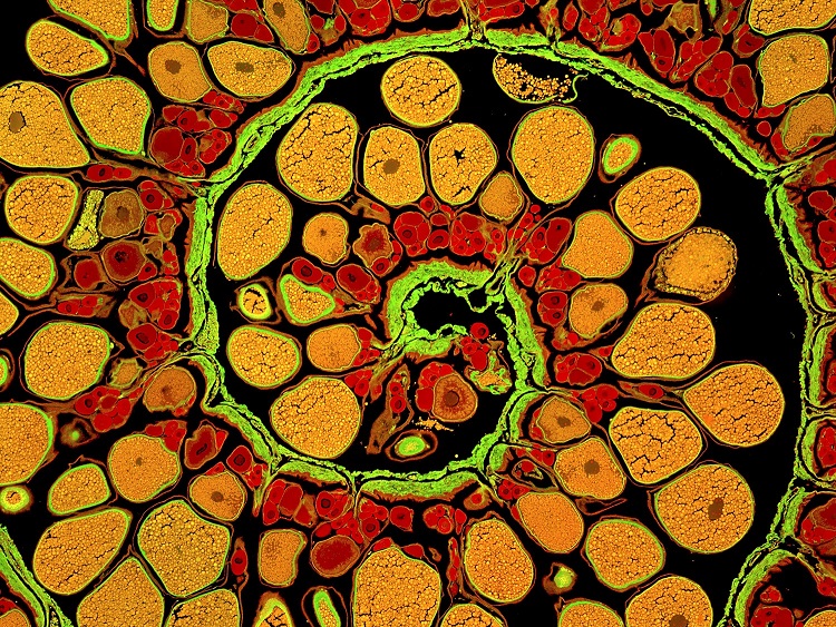
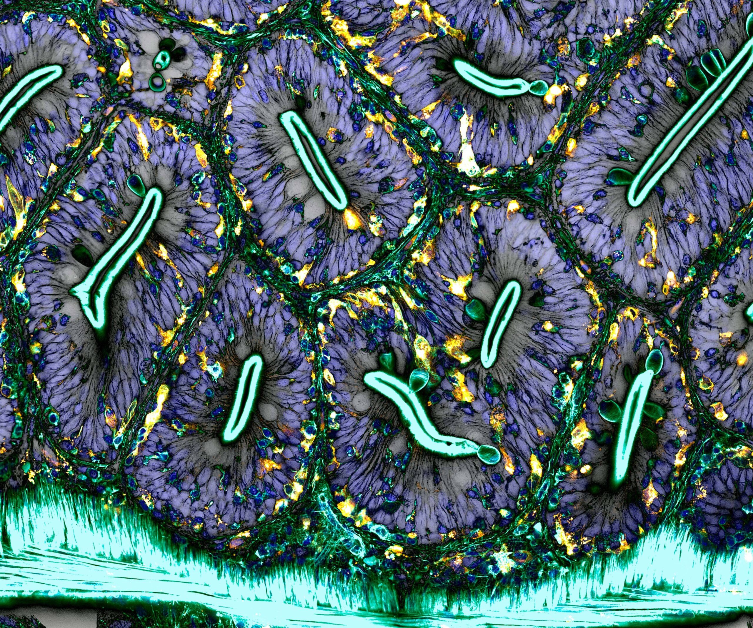
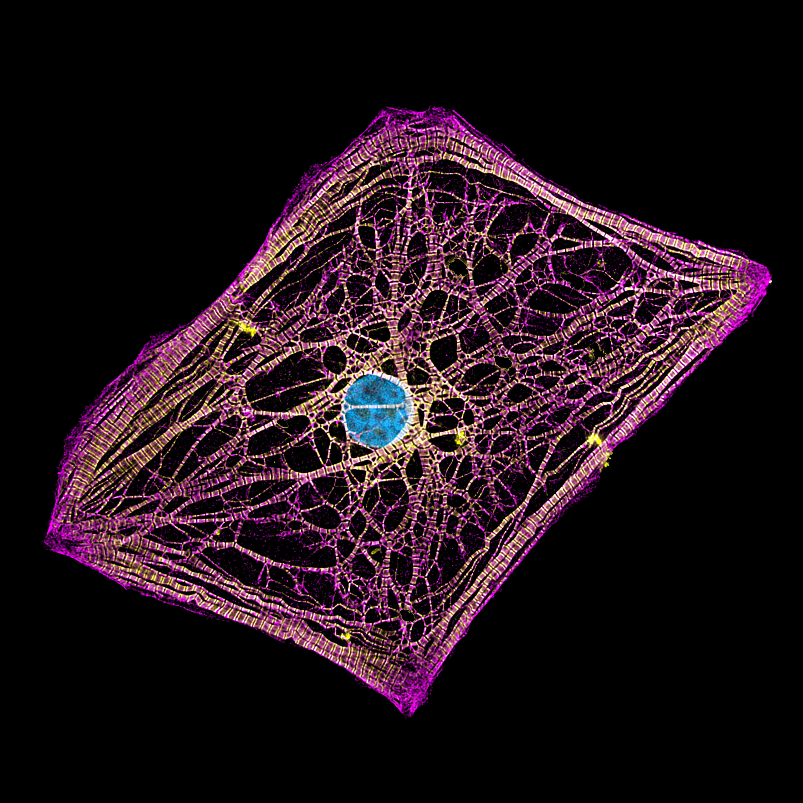
Breast Tissue: A Kaleidoscope of Life
Caleb Dawson, a dedicated researcher in the field of breast cancer, embraced the power of microscopy to uncover the mesmerizing intricacies of breast tissue. Through his lens, the once-invisible world of cellular composition comes alive with vibrant colors and fascinating structures. Within breast tissue, a tapestry of cells takes shape. Delicate clusters of glandular cells form breathtaking patterns, resembling an abstract painting. Ductal structures, reminiscent of branching trees, weave throughout the tissue. The microscopic exploration not only reveals the complexity of breast tissue but also underscores the importance of understanding its cellular landscape to advance medical research and treatments.
Emperor Butterfly: Nature’s Vibrant Brushstrokes
Renowned scientist Charles B. Krebs embarked on a journey into the microscopic world of the Emperor butterfly, captivated by its iridescent beauty. Through his microscope, the delicate wings of this majestic creature come to life, displaying a breathtaking palette of colors and intricate patterns. The butterfly’s wings, like stained-glass windows, showcase a mosaic of microscopic scales. Each scale, delicately arranged and overlapping, reflects and refracts light, creating a dazzling display of vibrant hues. The microscopic examination unravels the secrets behind nature’s mastery of color and reminds us of the endless wonders found in the animal kingdom.
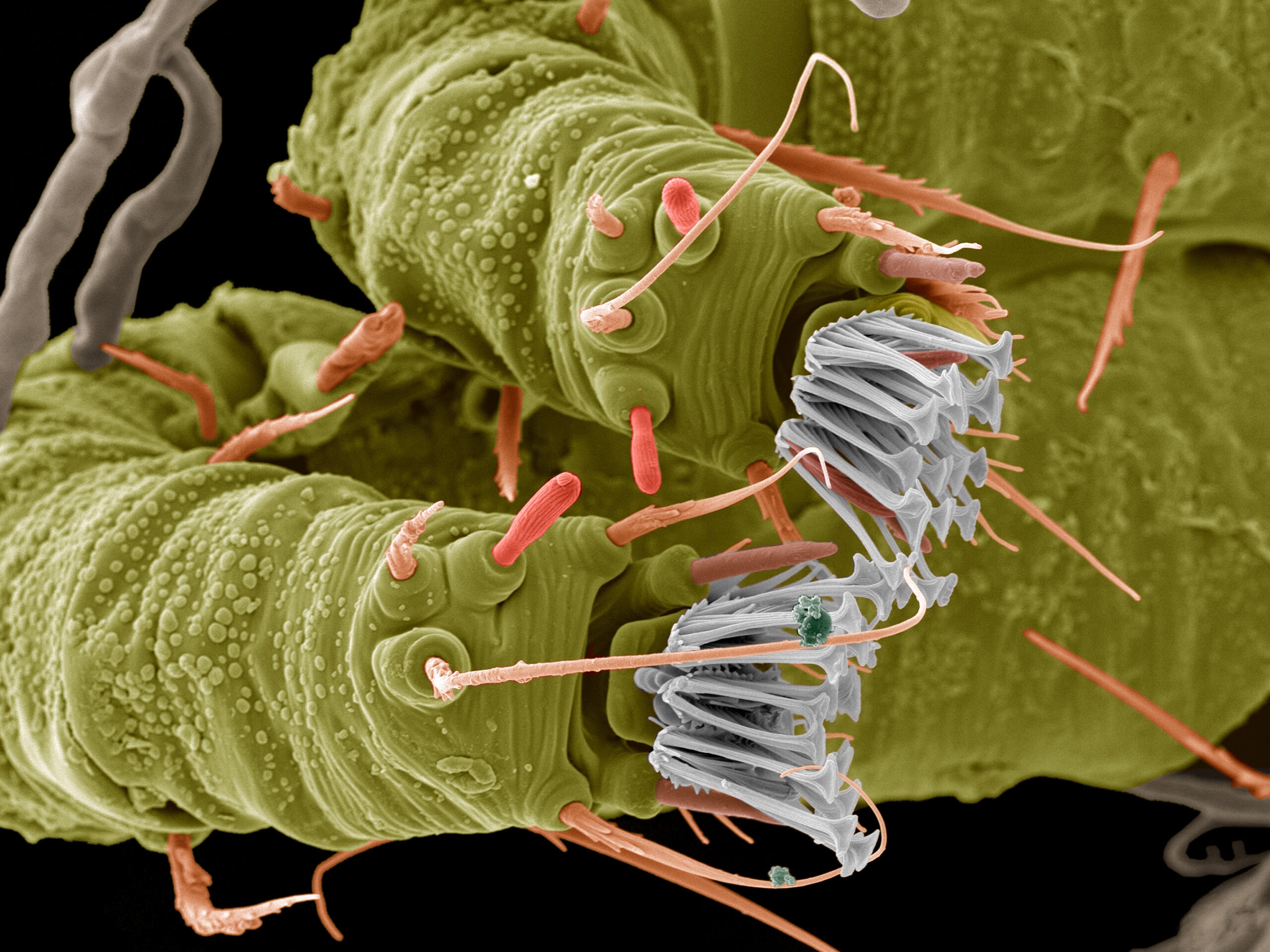
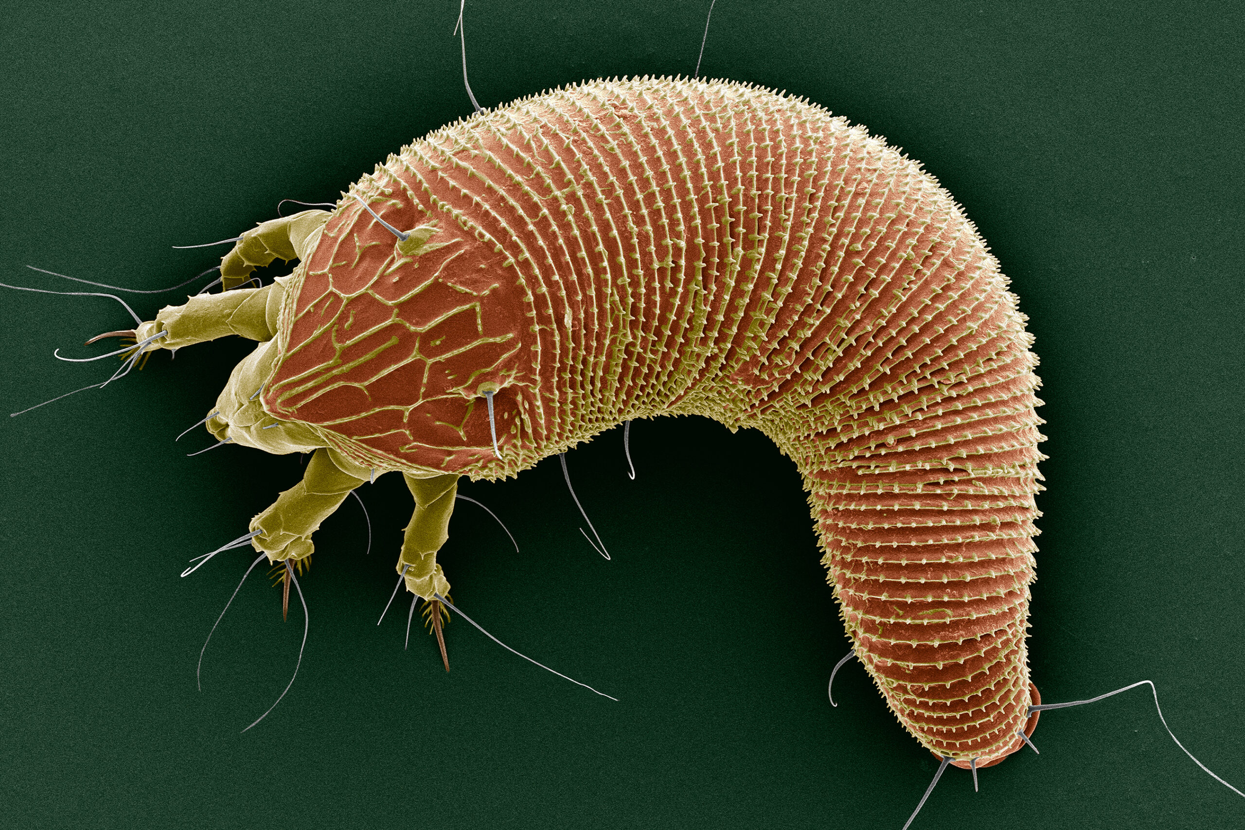
Mosquito Larva: A Portrait of Transformation
Anne Algar, a passionate entomologist, turned her microscope’s eye towards the mosquito larva, uncovering the hidden beauty within these often-maligned creatures. Through her lens, the larva’s aquatic world is transformed into a captivating symphony of shapes and textures. As we peer into the microscopic realm, the mosquito larva appears like a mesmerizing alien creature. Delicate bristles, resembling tiny hairs, line its body, enabling it to navigate through water with graceful precision. The segmented body and intricate mouthparts hint at its remarkable ability to adapt and transform throughout its life cycle. This exploration serves as a reminder that even the smallest and most overlooked creatures hold a fascinating beauty of their own.

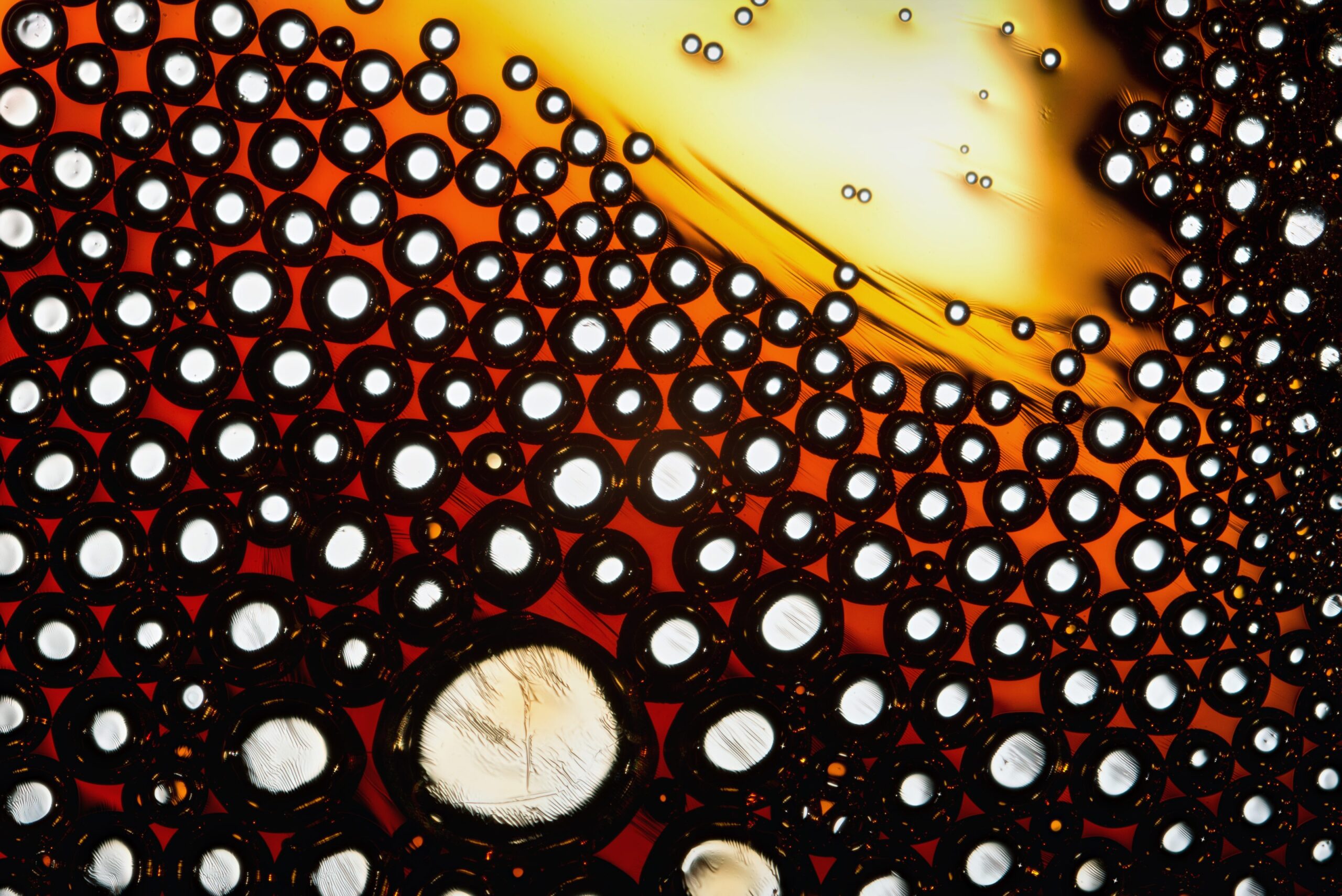
Molten Caffeine: An Intricate Dance of Molecules
Thomas Borowitz, an explorer of the chemical world, set his sights on the microscopic realm of molten caffeine. Through his microscope, the once-invisible dance of molecules reveals itself in a mesmerizing display of fluid dynamics. Molten caffeine, under extreme heat and magnification, presents a captivating spectacle. The individual molecules collide and intertwine, forming intricate patterns and trails. The molten caffeine dances with vibrant energy, its fluid nature shaping a dynamic and ever-changing landscape. The microscopic exploration captures the essence of molecular movement, offering a glimpse into the hidden beauty that lies within the world of chemistry.
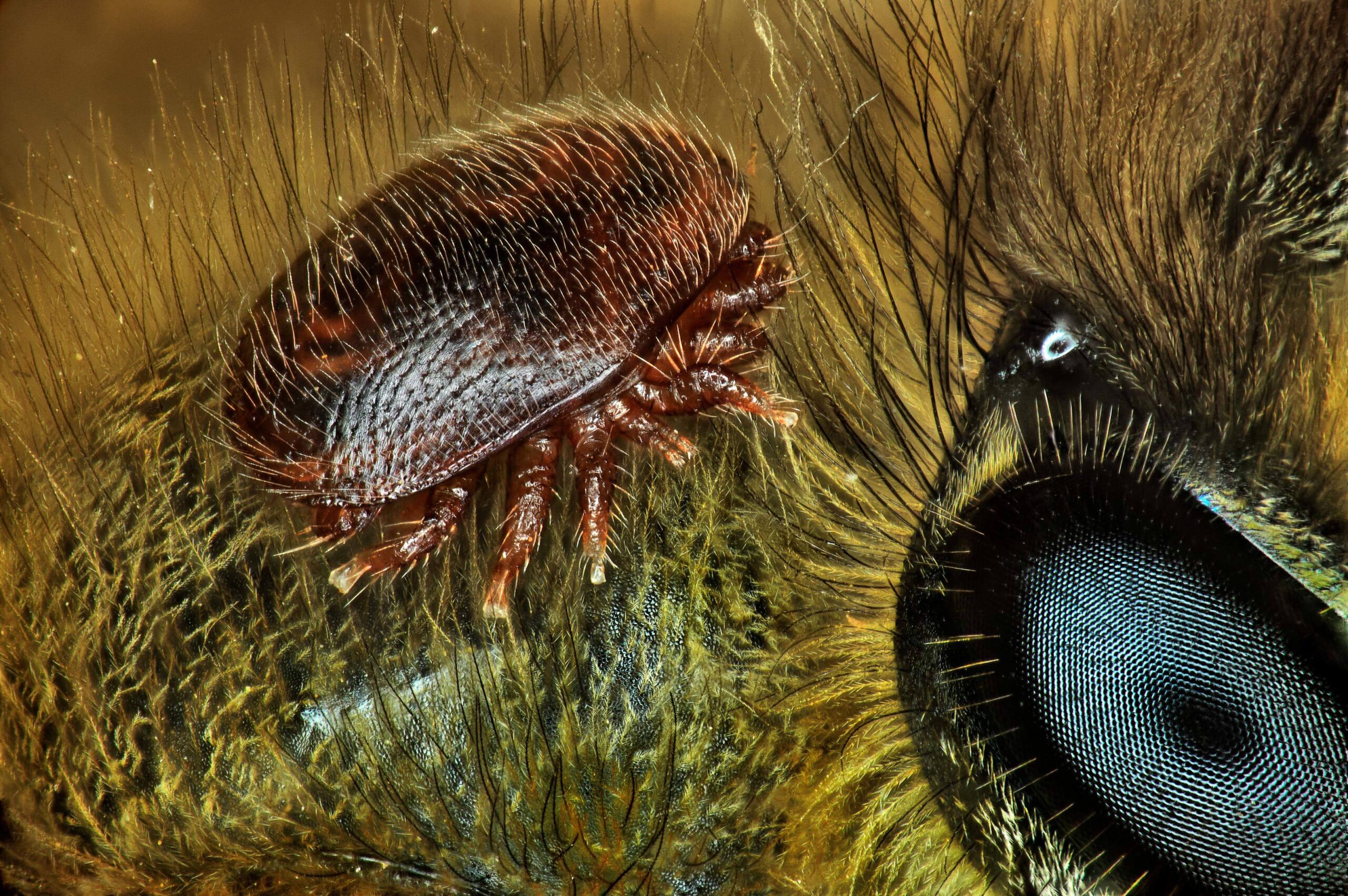
Mite-Honeybee: A Microscopic Partnership
Antoine Franck, a dedicated researcher and microscopy enthusiast, delved into the fascinating world of the mite-honeybee relationship. Through the lens of his microscope, Franck unraveled the intricate details of this symbiotic partnership that exists within beehives. As we delve into the microscopic world of a honeybee, we encounter a captivating sight. Tiny mites, known as Varroa destructor, cling to the bodies of bees, their translucent bodies and delicate appendages adding an ethereal touch to the scene. This microscopic exploration highlights the complexities of the relationship between mites and bees, shedding light on the delicate balance of nature and the challenges faced by these vital pollinators.
Mosses: A Microcosm of Delicate Beauty
Jacek Myslowski, a passionate botanist and microscopy enthusiast, embarked on a journey to uncover the hidden beauty within the world of mosses. Through his microscope, Myslowski revealed a microcosm of delicate beauty, captivating our senses with intricate structures and vibrant colors. As we peer into the microscopic realm of mosses, a new world unfolds before us. Tiny leaves, adorned with translucent cells and delicate structures, create a mesmerizing tapestry of shapes and patterns. The interplay of light and shadow adds depth to the scene, enhancing the ethereal beauty of these miniature plants. This microscopic exploration invites us to appreciate the often-overlooked wonders of the plant kingdom and reminds us of the intricate beauty that lies beneath our feet.
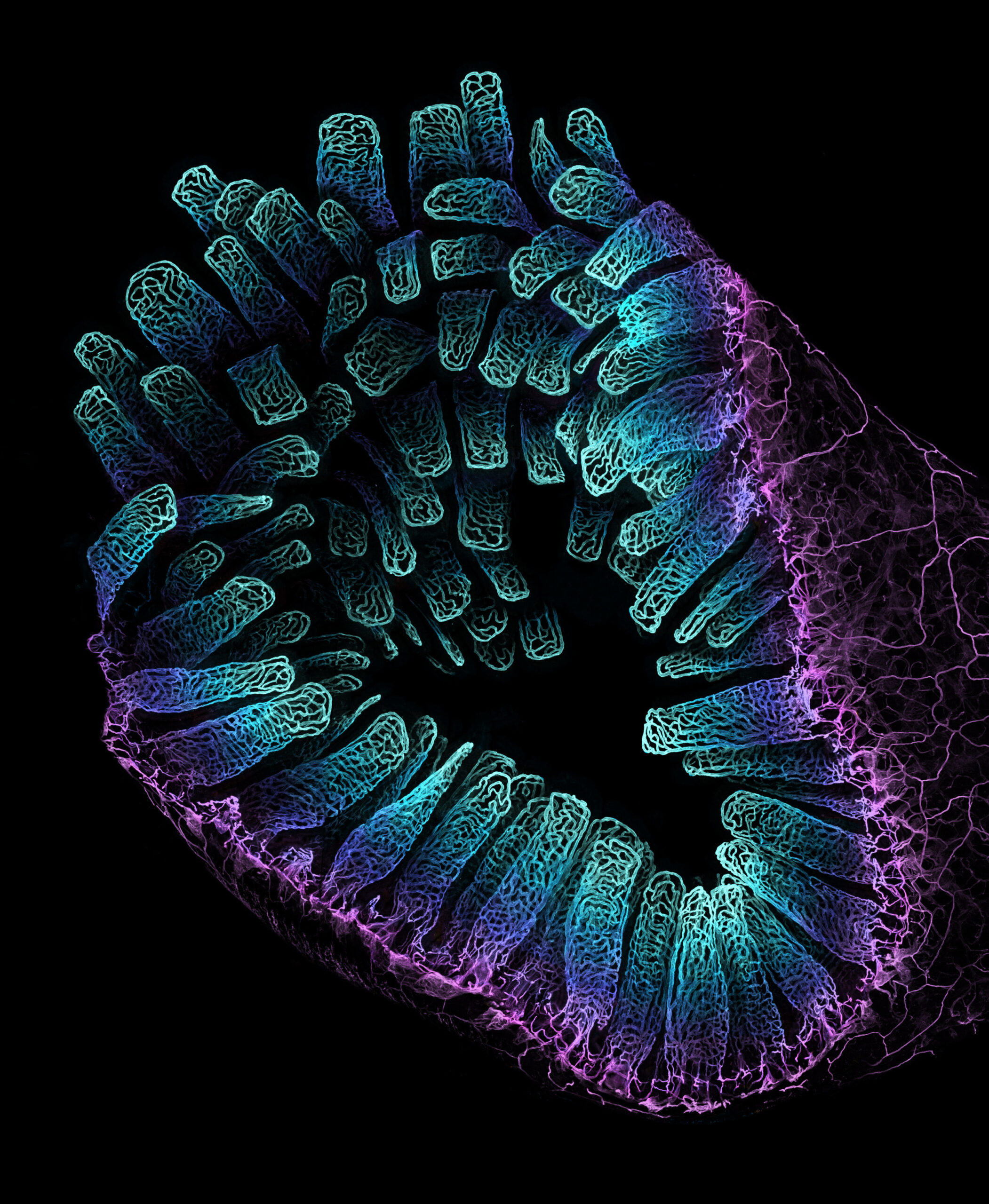
Zebrafish: A Symphony of Life
In the realm of developmental biology, Daniel Castranova, Dr. Brant M. Weinstein, and Bakary Samasa turned their attention to the microscopic world of zebrafish. Armed with their microscope, this team of researchers uncovered a captivating symphony of life unfolding within these tiny aquatic creatures. Under the microscope’s lens, zebrafish embryos reveal a breathtaking dance of growth and development. Transparent bodies showcase delicate organs and intricate blood vessels, pulsating with vibrant hues. The microscopic exploration not only offers insights into the fundamental processes of life but also highlights the astounding beauty found within these unassuming creatures. Through the lens of the microscope, we witness the wonders of biology unfolding in real-time.
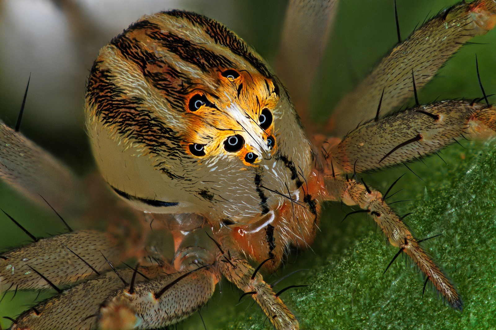
Female Lynx Spider: The Graceful Predator
Antoine Franck, with his insatiable curiosity, turned his microscope towards the world of female lynx spiders. In this microscopic exploration, Franck unraveled the hidden beauty within these small yet fierce hunters. As we observe the microscopic details of a female lynx spider, we are captivated by its elegant form and intricate features. The spider’s body is adorned with delicate hairs, each serving a specific purpose in its predatory lifestyle. The microscopic exploration reveals the astonishing adaptations that enable these spiders to thrive in their natural habitats, showcasing the beauty of nature’s design.
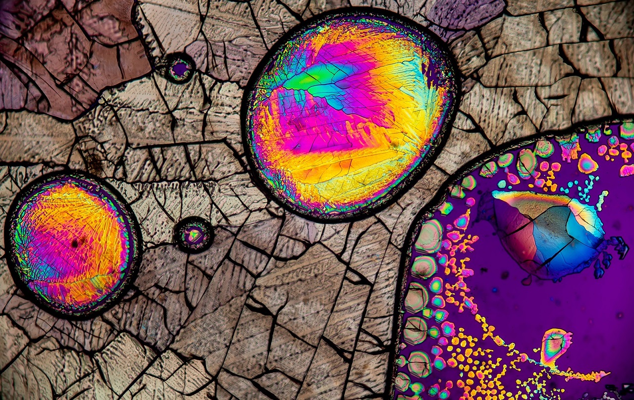
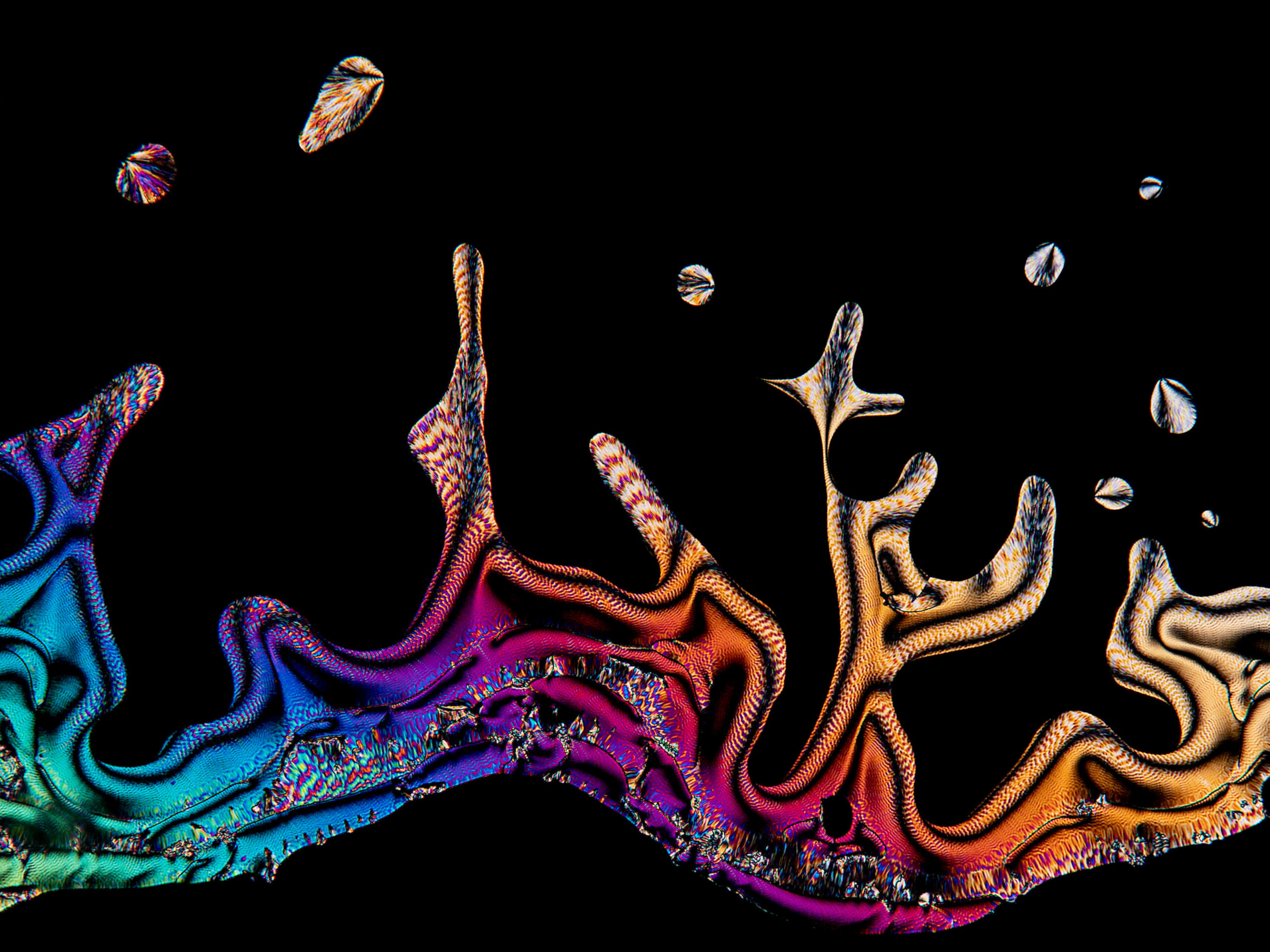
Amino Acid Crystals: The Building Blocks of Life
Justin Zoll, a passionate chemist and microscopy enthusiast, focused his attention on the crystalline world of amino acids. Through his microscope, Zoll revealed the intricate beauty that lies within these fundamental building blocks of life. A microscopic exploration of amino acid crystals unveils a symphony of geometric patterns and vibrant colors. Each crystal structure is a masterpiece of symmetry, with molecules aligning in intricate arrangements that give rise to stunning visual displays. The microscopic examination not only highlights the fundamental role of amino acids in life’s processes but also showcases the harmonious beauty of molecular architecture.
Mestra Butterfly Eggs on Tragia Leaf: A Tapestry of Life
Under the keen eye of David Millard, a passionate microscopy enthusiast, the microscopic world of Mestra butterfly eggs on a Tragia leaf is unveiled. Through his lens, we witness the delicate beauty and intricate complexity of these tiny eggs. As we peer into the microscopic realm, a tapestry of life unfolds before us. Each butterfly egg, resembling a tiny jewel, is meticulously attached to the leaf’s surface. The translucent shell of the egg reveals glimpses of the developing caterpillar within, reminding us of the miracles of life in its earliest stages. This microscopic exploration celebrates the remarkable process of reproduction and the delicate interplay between species.
Agatized Dinosaur Bone: Preserving Ancient Wonders
Randy Fullbright, a devoted researcher and microscopy enthusiast, turned his attention to the microscopic wonders locked within agatized dinosaur bone. Through his lens, Fullbright unraveled the intricate beauty preserved within these ancient remnants. Within the agatized dinosaur bone, a world of fossilized marvels awaits. Microscopic examination reveals intricate patterns and structures, reminiscent of a fossilized forest frozen in time. The interplay of vibrant colors and crystalline formations hints at the geological processes that shaped these magnificent remains over millions of years. This exploration not only captures the imagination but also deepens our understanding of Earth’s ancient past.
Bubonic Plague Bacteria: Unraveling a Historical Menace
ZEISS Microscopy, renowned for its cutting-edge imaging technology, provides us with a glimpse into the microscopic realm of bubonic plague bacteria. Through their advanced equipment, the intricate beauty and historical significance of these bacteria come to light. Under the microscope’s lens, we witness the hauntingly captivating form of the Yersinia pestis bacteria, the causative agent of the infamous bubonic plague. The rod-shaped bacteria intertwine, forming intricate patterns that both fascinate and remind us of the devastating impact of this historical disease. This microscopic exploration serves as a testament to the delicate balance between life and death and the ongoing battle between humanity and infectious diseases.
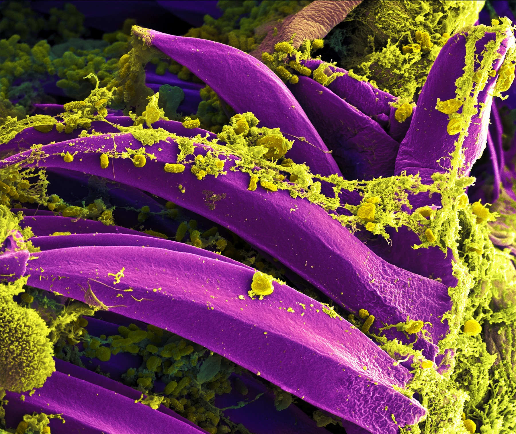
Daddy Long-leg Eye: A Window to the World
Charles B. Krebs, a visionary in the realm of microscopy, turns his gaze towards the intricate eyes of a daddy long-leg spider. Through his lens, Krebs unveils the hidden beauty and remarkable visual adaptations of these fascinating creatures. As we delve into the microscopic world of the daddy long-leg, the intricacies of its compound eyes come into focus. Delicate structures, resembling a mosaic of tiny lenses, provide the spider with a panoramic view of its surroundings. The microscopic exploration not only showcases the marvels of nature’s engineering but also invites us to appreciate the intricate visual systems found within the animal kingdom.
Beetle: Nature’s Architectural Marvel
ZEISS Microscopy, with its powerful imaging capabilities, takes us on a microscopic journey into the world of beetles. Through their lens, we discover the astounding artistry and architectural wonders of these tiny creatures. Under the microscope, the exoskeleton of a beetle reveals a breathtaking display of intricate structures and textures. Each tiny ridge and groove plays a vital role in the beetle’s survival and adaptation. The microscopic exploration unravels the secrets of nature’s craftsmanship, showcasing the beetle’s remarkable ability to create such intricate and durable armor. From the mesmerizing patterns on its wings to the intricate texture of its exoskeleton, the beetle becomes a testament to the beauty and ingenuity found within the natural world.
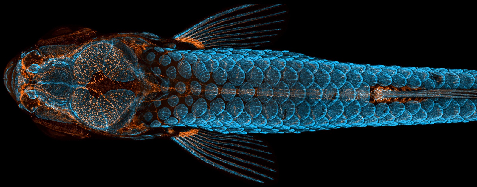
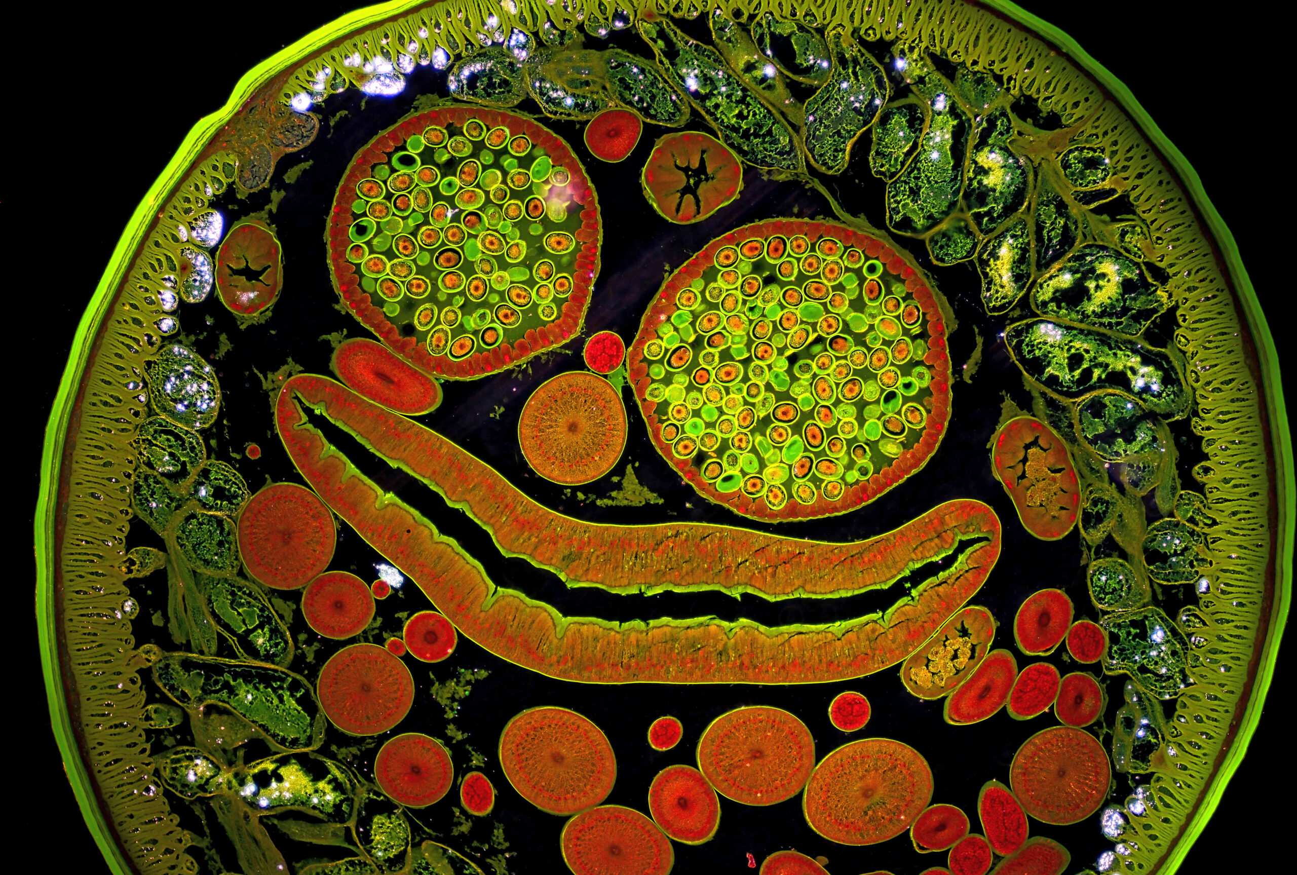
Angelfish Ovary: Nature’s Masterpiece
ZEISS Microscopy, renowned for its cutting-edge imaging technology, invites us to peer into the microscopic world of an angelfish ovary. Through their advanced equipment, the intricate beauty and remarkable complexity of this reproductive organ are brought to light. Under the microscope’s lens, the angelfish ovary unveils a breathtaking symphony of vibrant colors and delicate structures. Each egg, poised for potential new life, is surrounded by a delicate network of cells and blood vessels. This microscopic exploration not only celebrates the wonders of reproduction but also reveals the stunning artistry inherent in nature’s design.
Red Algae: A Kaleidoscope of Marine Life
In the hidden depths of our oceans, a microscopic spectacle unfolds as we delve into the world of red algae. Through the lens of a microscope, we discover an enchanting display of colors, shapes, and intricate patterns. As we peer into the microscopic realm, we are captivated by the beauty of red algae. Delicate filaments intertwine, forming an otherworldly tapestry of forms and textures. The vibrant reds, oranges, and greens create a mesmerizing kaleidoscope of marine life, reminding us of the immense biodiversity that thrives beneath the ocean’s surface.
Dyed Human Hair: A Playground of Colors
Harald K. Andersen, a visionary in microscopy, directs his lens towards the microscopic wonders found within dyed human hair. Through his artistic exploration, Andersen unravels a hidden world of vibrant colors and fascinating structures. Under the microscope’s gaze, dyed human hair reveals a stunning spectrum of colors and intricate patterns. The individual strands become a playground for artistic expression, showcasing a tapestry of vibrant hues and dynamic textures. This microscopic exploration celebrates the fusion of human creativity and the natural beauty of our own bodies.
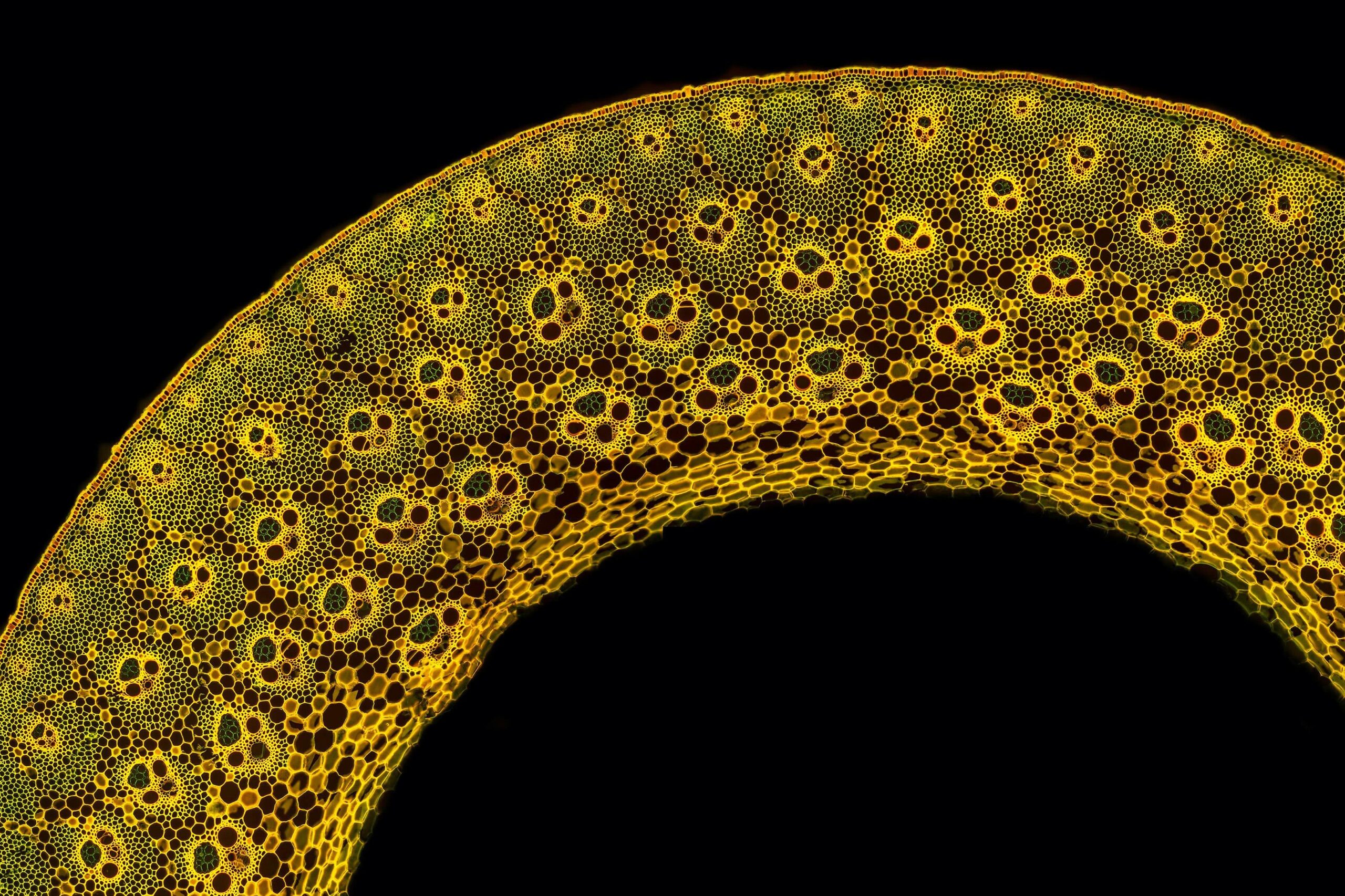
Garden Bamboo: Nature’s Poetic Architecture
Gerd Günther, a passionate microscopy enthusiast, directs our attention to the hidden beauty within the world of garden bamboo. Through his lens, Günther unveils the poetic architecture and intricate details of this versatile plant. As we delve into the microscopic world of garden bamboo, we witness the intricate structure of its cells and fibers. The interplay of light and shadow highlights the elegant and rhythmic patterns that make up the plant’s composition. This microscopic exploration invites us to appreciate the delicate balance between strength and flexibility, reminding us of nature’s remarkable engineering.
Blood Vessel Networks: Lifelines Within
Satu Paavonsalo and Dr. Sinem Karaman, pioneers in vascular research, turn their attention to the microscopic wonders of blood vessel networks. Through their advanced imaging techniques, they uncover the hidden beauty and intricate complexity of our circulatory system. Under the microscope, blood vessel networks reveal a breathtaking display of branching pathways and interconnectedness. The delicate capillaries, like fragile threads, carry life-sustaining oxygen and nutrients throughout our bodies. This microscopic exploration not only deepens our understanding of the intricate machinery that keeps us alive but also celebrates the miraculous beauty found within the human body.
Eyeshadow Cosmetic: A Kaleidoscope of Colors
Teresa Zgoda, a visionary in the world of microscopy, turns her attention to the microscopic wonders found within eyeshadow cosmetics. Through her lens, Zgoda unveils a mesmerizing world of vibrant pigments and intricate textures. As we peer into the microscopic realm, the eyeshadow cosmetic transforms into a kaleidoscope of colors. Delicate particles dance and intertwine, creating a visual symphony of shimmering hues and dazzling reflections. This microscopic exploration celebrates the artistry of makeup and the power of self-expression through color and texture.
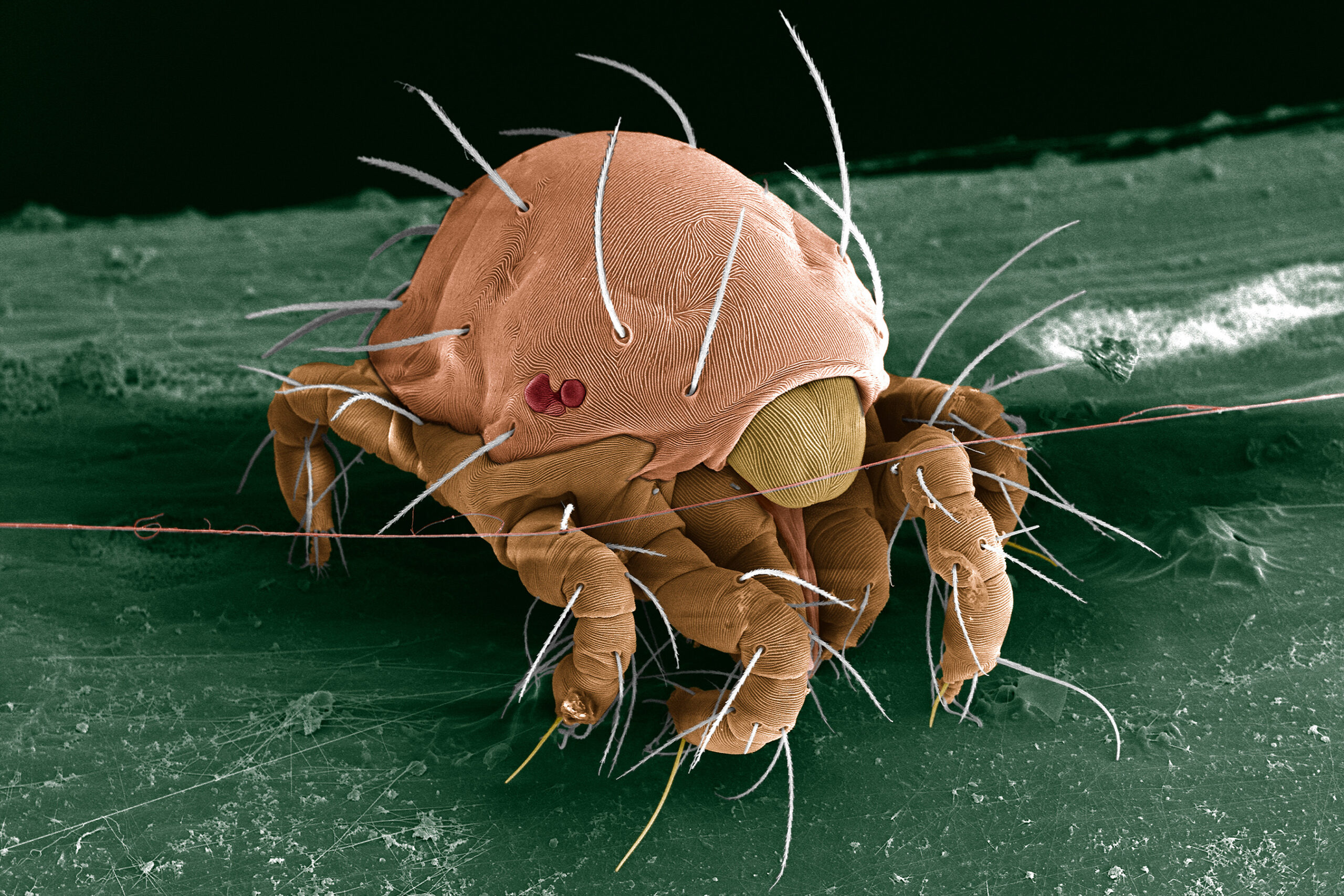
Mite Front Legs: Nature’s Acrobats
In the hidden world of mites, microscopic acrobats await our discovery. Through the lens of a microscope, we delve into the intricate details of mite front legs, unraveling a world of astonishing adaptations. As we examine the microscopic structure of mite front legs, we witness an awe-inspiring display of complexity. Tiny hairs, hooks, and claws adorn the legs, allowing these microscopic creatures to navigate their environment with astonishing precision. This exploration showcases the marvels of nature’s engineering and reminds us of the remarkable adaptations that enable life to thrive in even the most challenging conditions.
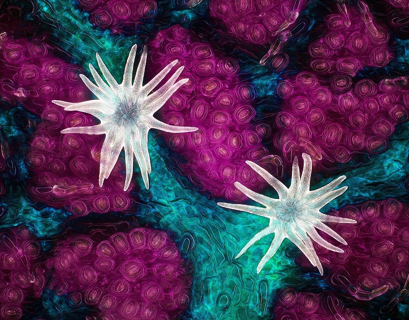
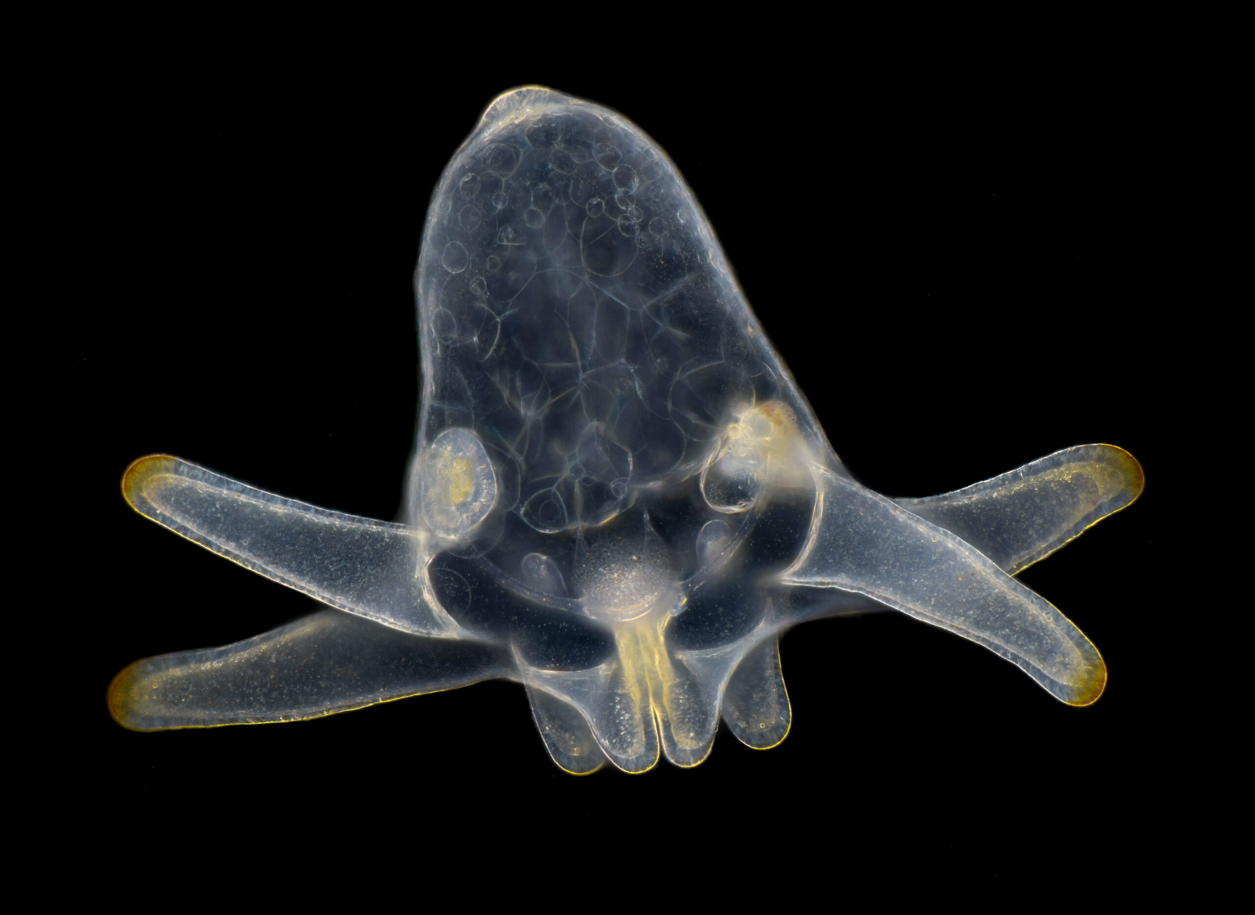
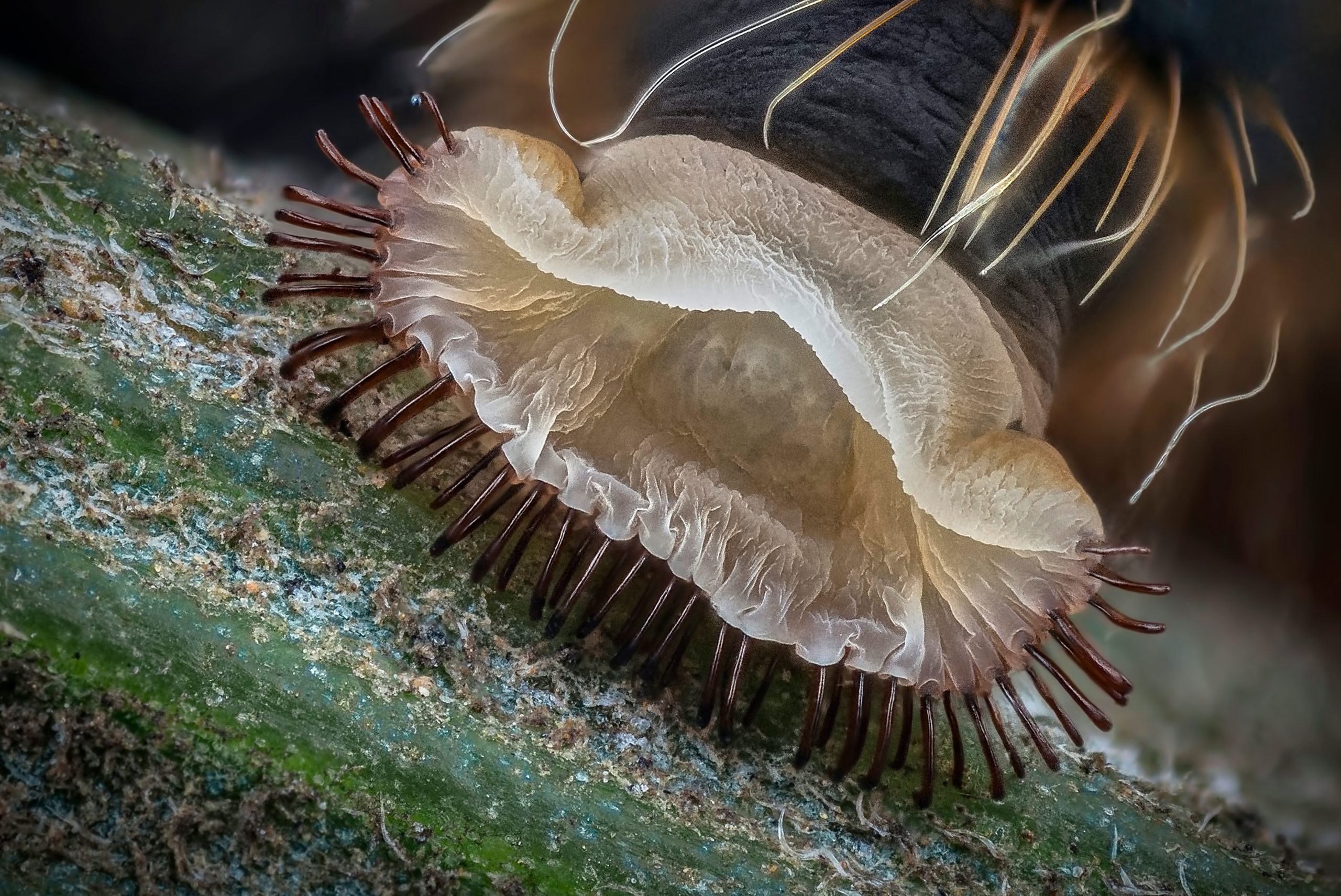
Larva of Anemone: A Delicate Journey
Wim van Egmond, a passionate microscopy enthusiast, directs our attention to the hidden beauty within the larva of anemone. Through his lens, van Egmond unveils the delicate transformation and intricate details of this stage in the life cycle of an anemone. Under the microscope, the larva of anemone reveals a mesmerizing display of transparent structures and intricate patterns. Delicate tentacles and cilia come to life, showcasing the larva’s ability to move and feed. This microscopic exploration captures the fragility and resilience of life, reminding us of the wonder and beauty that exist within the hidden stages of natural processes.
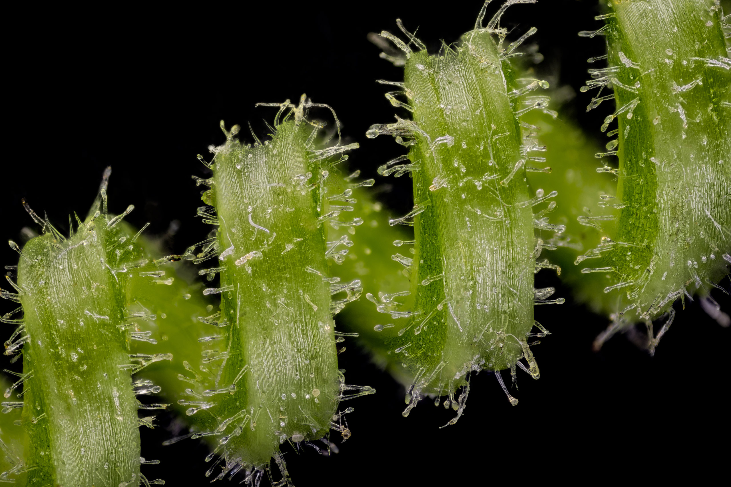
Dune Grass Leaf: A Symphony of Texture
Anatoly Mikhaltsov, a visionary in the world of microscopy, takes us on a journey into the microscopic world of a dune grass leaf. Through his lens, Mikhaltsov reveals the intricate textures and patterns that adorn this seemingly simple leaf. As we delve into the microscopic realm of the dune grass leaf, a symphony of texture unfolds. Delicate hairs, intricate veins, and unique cellular structures create a visually captivating landscape. This exploration invites us to appreciate the beauty of even the simplest elements of nature, showcasing the intricate details that exist within every living organism.
Housefly: Nature’s Unsung Hero
Oliver Dum, a dedicated microscopy enthusiast, directs our gaze towards the microscopic wonders of a housefly. Through his lens, Dum unveils the hidden beauty and remarkable adaptations of this often-overlooked insect. Under the microscope, the housefly transforms into a world of intricate structures and delicate patterns. From the compound eyes that provide a panoramic view of its surroundings to the intricate bristles and sensory organs that adorn its body, the housefly showcases the remarkable beauty and resilience of nature’s creations. This microscopic exploration invites us to look beyond our preconceived notions and appreciate the often-unseen wonders that surround us.
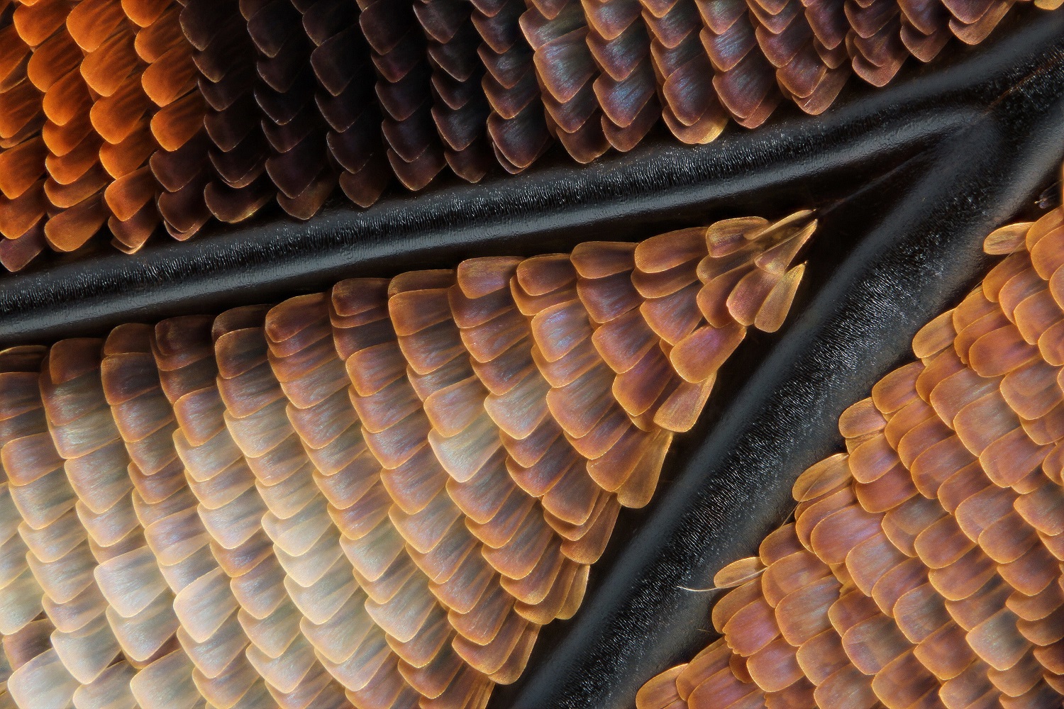
Butterfly Wing: Nature’s Living Canvas
Francis Sneyers, a master of microscopy, guides us into the captivating world of butterfly wings. Through his lens, Sneyers reveals the exquisite beauty and intricate patterns that adorn these delicate structures. As we delve into the microscopic realm of butterfly wings, a world of mesmerizing colors and patterns unfolds before us. Each tiny scale, like a brushstroke on a canvas, contributes to a symphony of hues and designs. From iridescent blues and vibrant oranges to delicate patterns that mimic the wonders of nature, butterfly wings are a testament to the artistry found in every corner of the natural world.
Freshwater Snail: Nature’s Architect
Dr. Igor Siwanowicz, a renowned microscopy expert, invites us to explore the microscopic wonders of freshwater snails. Through his lens, Siwanowicz uncovers the intricacies of their shells and the fascinating adaptations that make these mollusks true marvels of nature. Under the microscope, the shells of freshwater snails become a masterpiece of engineering and design. Intricate spirals, delicate ridges, and mesmerizing patterns create a visually stunning display. This microscopic exploration not only celebrates the beauty of these tiny creatures but also reminds us of the remarkable diversity and ingenuity found in the natural world.
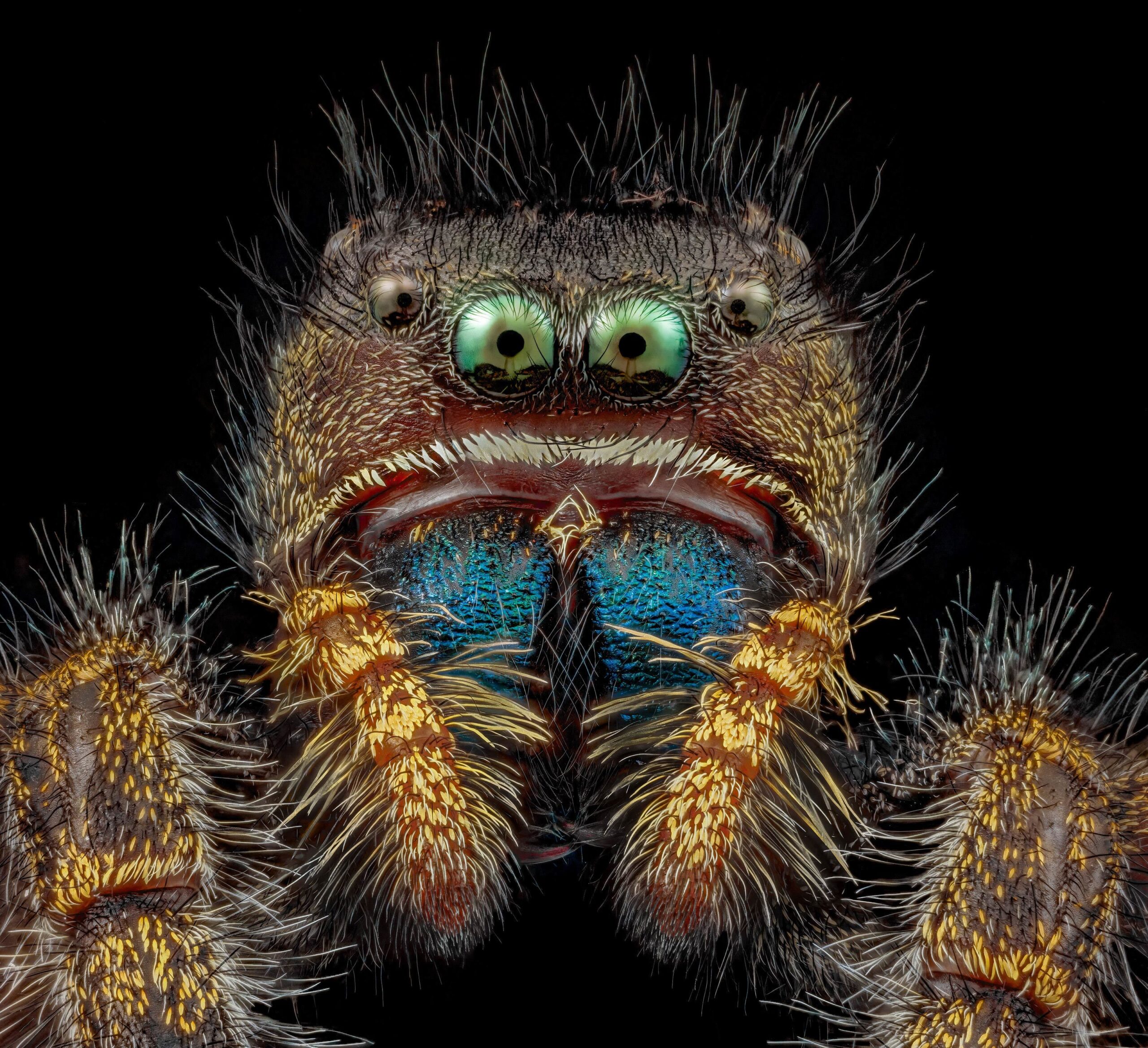
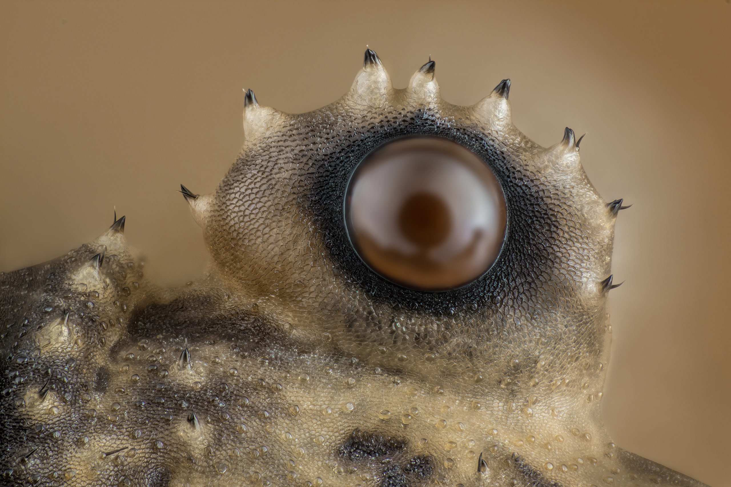
Jumping Spider: Eyes of a Hunter
Emre Can Alagöz, a passionate microscopy enthusiast, focuses his lens on the intricate eyes of a jumping spider. Through his exploration, Alagöz unveils the remarkable adaptations that make these tiny predators such formidable hunters. As we peer into the microscopic world of jumping spider eyes, we discover an array of optical wonders. Multiple pairs of eyes, each with their own unique capabilities, grant the spider a 360-degree view of its surroundings. The microscopic details, from the arrangement of lenses to the sensitivity of light receptors, highlight the spider’s extraordinary visual prowess. This exploration showcases the intricate beauty and extraordinary adaptations that enable these spiders to navigate their world with precision and agility.
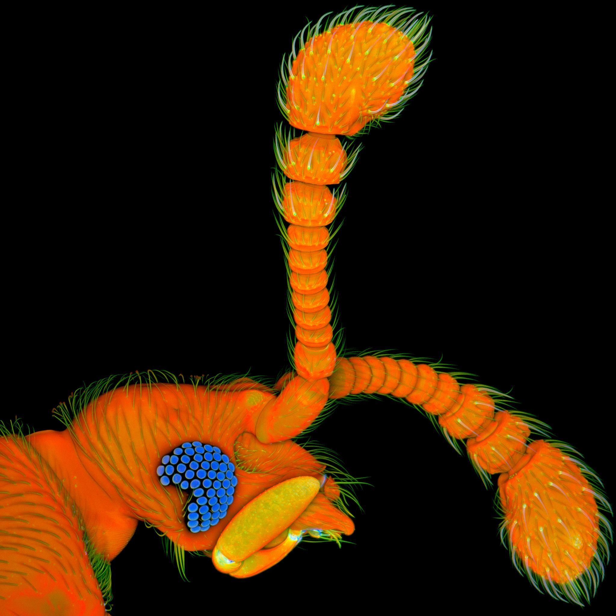
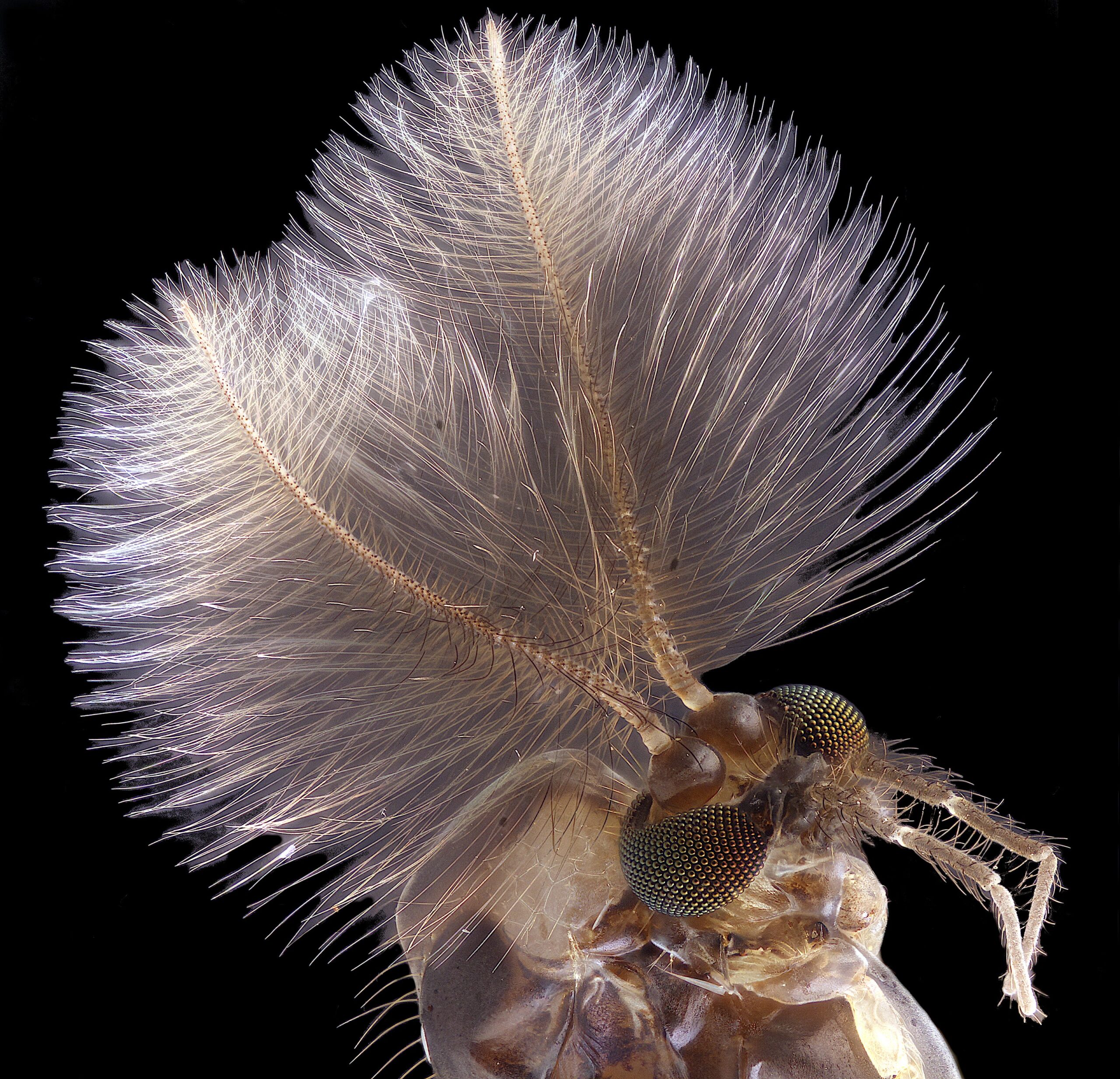
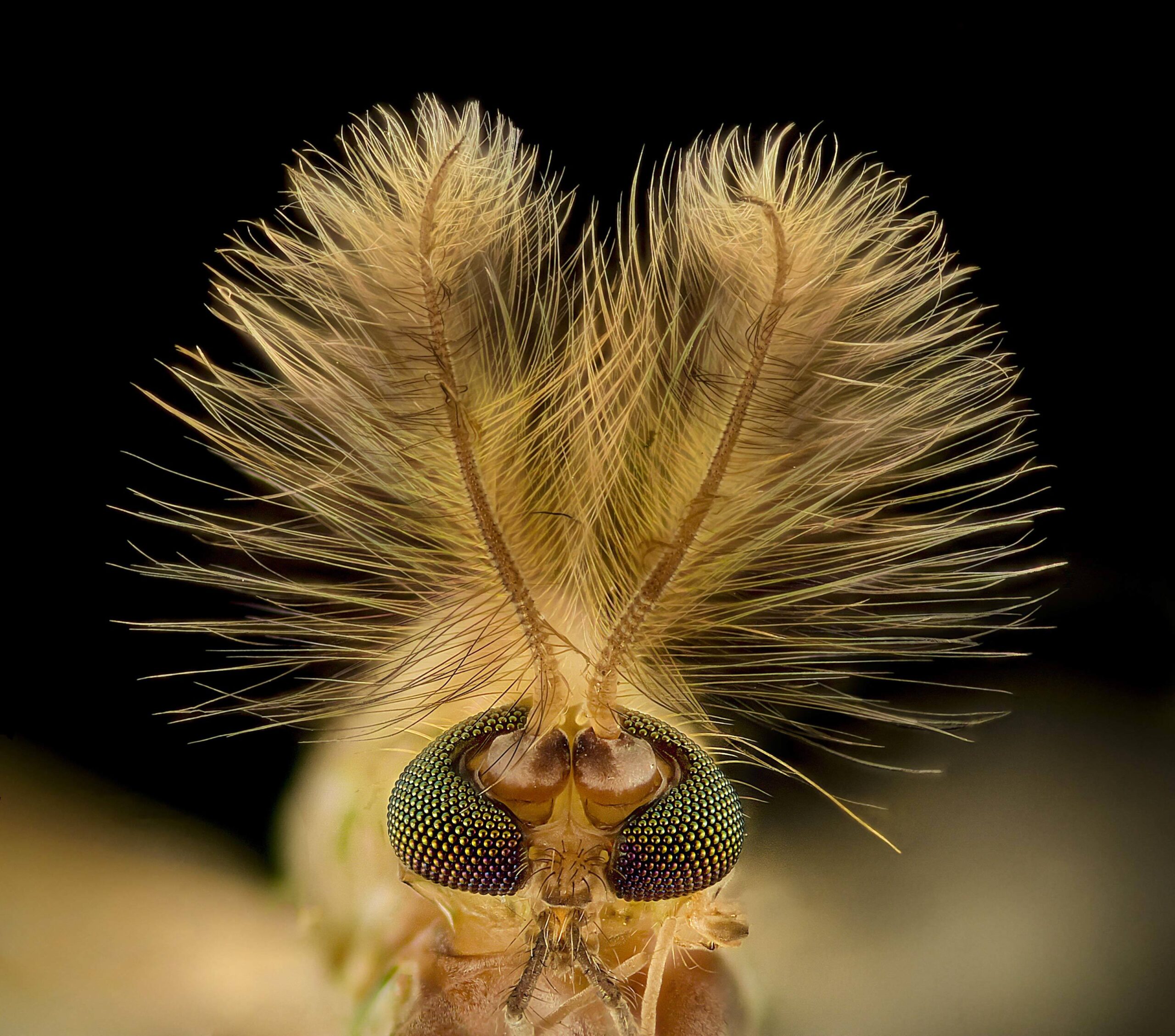
Male Mosquito: Beauty in the Tiny Details
Jan Rosenboom, an esteemed microscopy expert, directs our attention to the microscopic wonders found within the male mosquito. Through his lens, Rosenboom reveals the hidden beauty and intricate structures that define these often-maligned insects. Under the microscope, the male mosquito transforms into a delicate marvel of nature. Intricate mouthparts, intricate antennae, and fine hairs become visible, showcasing the mosquito’s remarkable adaptations for survival and reproduction. This exploration reminds us that even in the smallest and most overlooked creatures, beauty and complexity can be found.
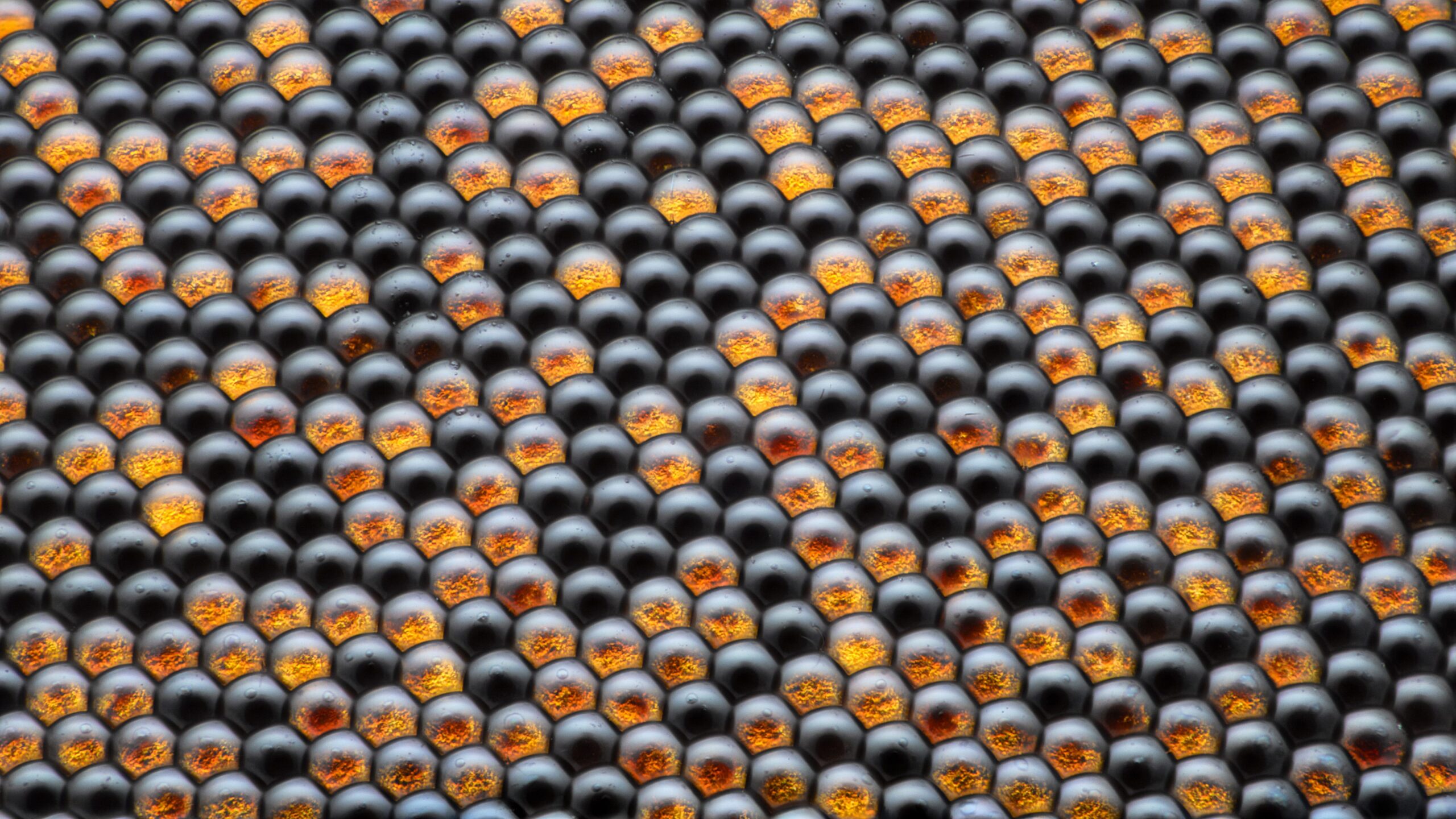
Honey Bee Eye: The World in a Hexagon
Ralph Grimm, a visionary in the world of microscopy, invites us to gaze into the eyes of a honey bee. Through his lens, Grimm unravels the hidden beauty and fascinating structure of the compound eyes that define these industrious insects. Under the microscope, the honey bee’s compound eyes become a mesmerizing mosaic of hexagonal units. Each facet captures a unique perspective of the world, creating a remarkable composite image. The microscopic exploration of the honey bee’s eye not only celebrates the intricate beauty of this essential pollinator but also reminds us of the wonder and complexity of nature’s creations.
Crystals: Nature’s Prismatic Gems
Justin Zoll, a master of microscopy, transports us into the enchanting world of crystals. Through his lens, Zoll unveils the mesmerizing beauty and intricate structures that lie within these geological wonders. Under the microscope, crystals transform into a kaleidoscope of dazzling colors and geometric patterns. Delicate facets and symmetrical arrangements create a visual feast for the eyes. From the sharp edges of quartz to the delicate formations of snowflakes, crystals showcase the natural artistry that emerges from the interplay of molecular forces. This microscopic exploration invites us to marvel at the harmonious beauty found in the smallest building blocks of our planet.

Beetle Leg: A Microcosm of Adaptation
Aigars Jukna, a passionate microscopy enthusiast, turns our attention to the microscopic wonders of a beetle leg. Through his lens, Jukna unravels the remarkable adaptations and intricate structures that make these insect appendages a true marvel of nature. As we delve into the microscopic world of a beetle leg, we are captivated by its complex and functional design. Fine hairs, sturdy exoskeletons, and intricate joints work in perfect harmony, enabling these tiny creatures to navigate their surroundings with astonishing agility and strength. This exploration celebrates the ingenuity of nature’s engineering, reminding us of the incredible adaptations that allow life to thrive in diverse environments.
White Sugar: Crystalline Sweetness
ZEISS Microscopy, renowned for its advanced imaging technology, invites us to peer into the microscopic world of white sugar. Through their powerful microscopes, they unravel the hidden beauty and unique structures that lie within this beloved culinary ingredient. Under the microscope, white sugar crystals dazzle with their delicate and intricate formations. Each crystal, composed of countless individual sugar molecules, comes to life as a testament to the beauty and complexity found in even the simplest of substances. This microscopic exploration reminds us that beauty can be found in unexpected places, even in the ingredients we use in our everyday lives.
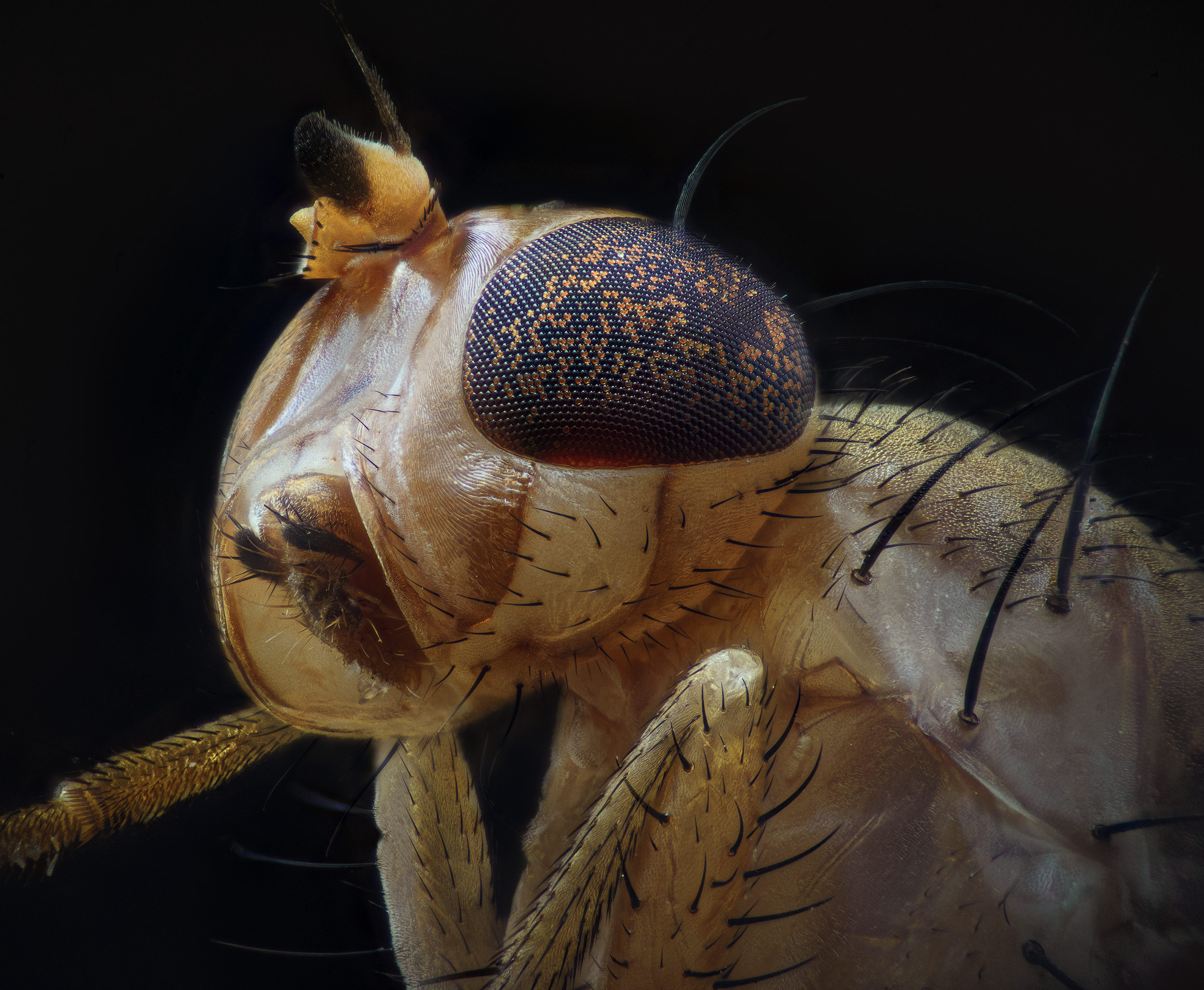
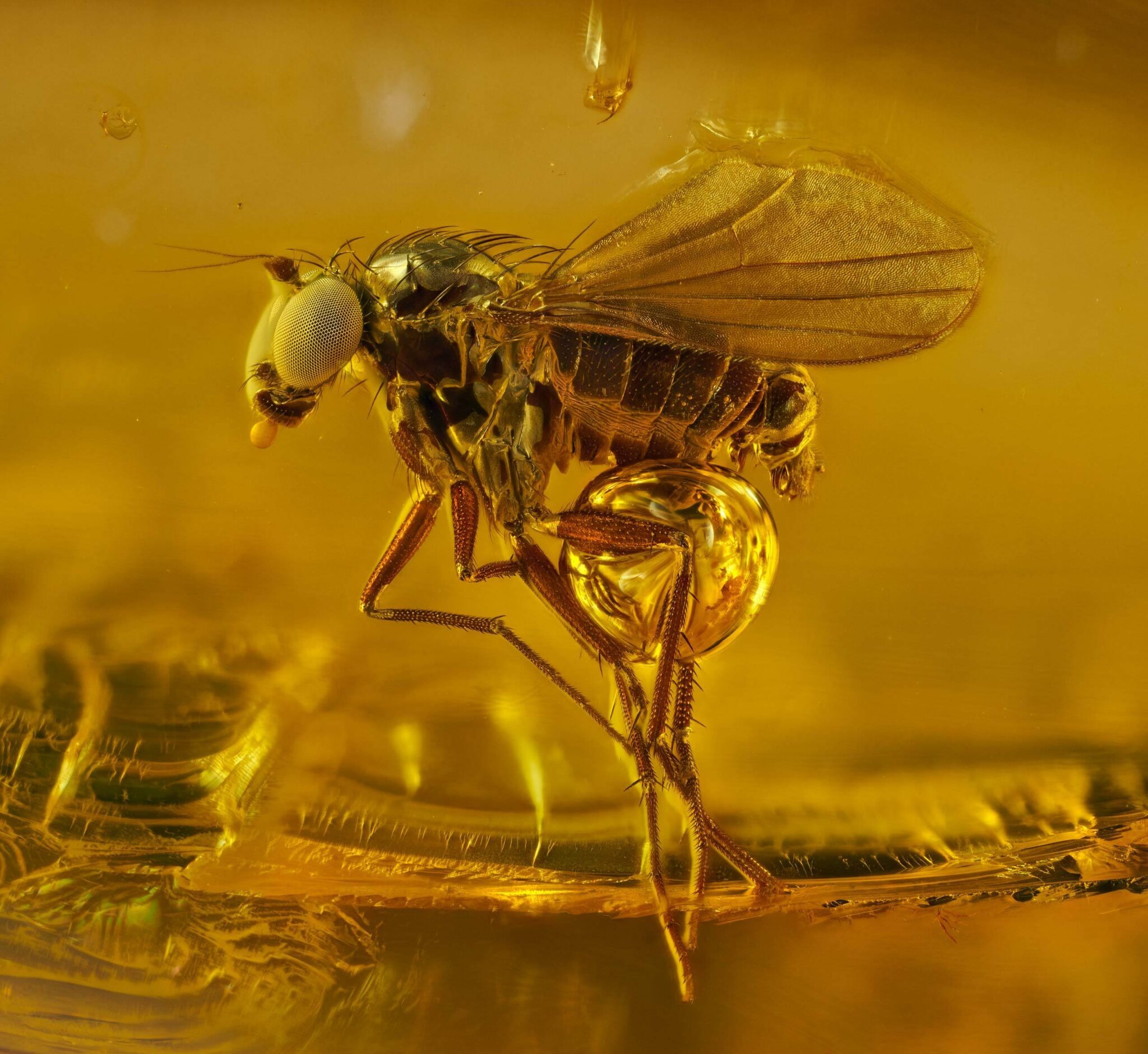
Vinegar Fly: Wings of Elegance
In the hidden realm of vinegar flies, microscopic wonders await our discovery. Through the lens of a microscope, we unveil the delicate beauty of their transparent wings, which allow these tiny insects to flutter through the air with grace. As we examine the microscopic details of vinegar fly wings, we are captivated by their intricate structure. Fine veins, delicate hairs, and unique patterns adorn the wings, reflecting the elegance and functionality of this essential appendage. This exploration reminds us of the hidden marvels that exist within the tiniest creatures, inviting us to appreciate the beauty and diversity that surround us.
Vitamin C: Microscopic Nutrient Powerhouse
Sebastian Sparenga, a visionary in microscopy, directs our attention to the microscopic wonders of vitamin C. Through his lens, Sparenga uncovers the hidden beauty and intricate structures that define this essential nutrient. Under the microscope, vitamin C crystals reveal a fascinating array of formations and shapes. The intricate lattice structure of these crystals showcases the molecular arrangement that gives vitamin C its powerful antioxidant properties. This microscopic exploration not only celebrates the beauty of this vital nutrient but also highlights the importance of microscopic elements in our overall health and well-being.
Sea Cucumber Skin: A Textured Marine Tapestry
Christian Gautier, a skilled microscopy artist, takes us on a journey into the microscopic realm of sea cucumber skin. Through his lens, Gautier reveals the intricate textures and patterns that adorn these fascinating marine creatures. As we peer into the microscopic world of sea cucumber skin, a tapestry of textures unfolds. Delicate ridges, spiky projections, and intricate patterns create a visually captivating landscape. This exploration invites us to appreciate the remarkable diversity and beauty that exists within the hidden corners of the ocean.
Slime Mold: Nature’s Intricate Network
Alison Pollack, a passionate microscopy enthusiast, directs our attention to the hidden wonders of slime mold. Through her lens, Pollack unveils the intricate and mesmerizing structures that make up these unique organisms. Under the microscope, slime mold reveals a sprawling network of delicate filaments and interconnected pathways. The microscopic exploration showcases the adaptive abilities of slime mold, as it creates complex branching structures to optimize nutrient absorption and reproduction. This exploration reminds us of the resilience and resourcefulness of nature’s creations, even in the most unexpected forms.
Bindweed Leaf: The Elegance of Nature’s Patterns
Michael Gibson, a visionary in the world of microscopy, focuses his lens on the intricate beauty of a bindweed leaf. Through his exploration, Gibson unravels the hidden patterns and delicate structures that define this common plant. Under the microscope, the bindweed leaf transforms into an artistic masterpiece of intricate veins and graceful patterns. Each tiny detail, meticulously arranged, contributes to the overall elegance and functionality of the leaf. This microscopic journey celebrates the natural artistry that exists within even the simplest elements of the botanical world.
Digger Wasp Eyes: Precision in Vision
Laurie Knight, an esteemed microscopy expert, directs our gaze towards the microscopic wonders found within the eyes of a digger wasp. Through her lens, Knight unveils the hidden beauty and remarkable adaptations of these tiny insect eyes. Under the microscope, the digger wasp’s eyes reveal an intricate array of lenses and receptors, providing these insects with remarkable visual acuity. The microscopic exploration highlights the precision and sophistication of nature’s designs, as the eyes of digger wasps allow them to navigate their surroundings with incredible precision and accuracy.
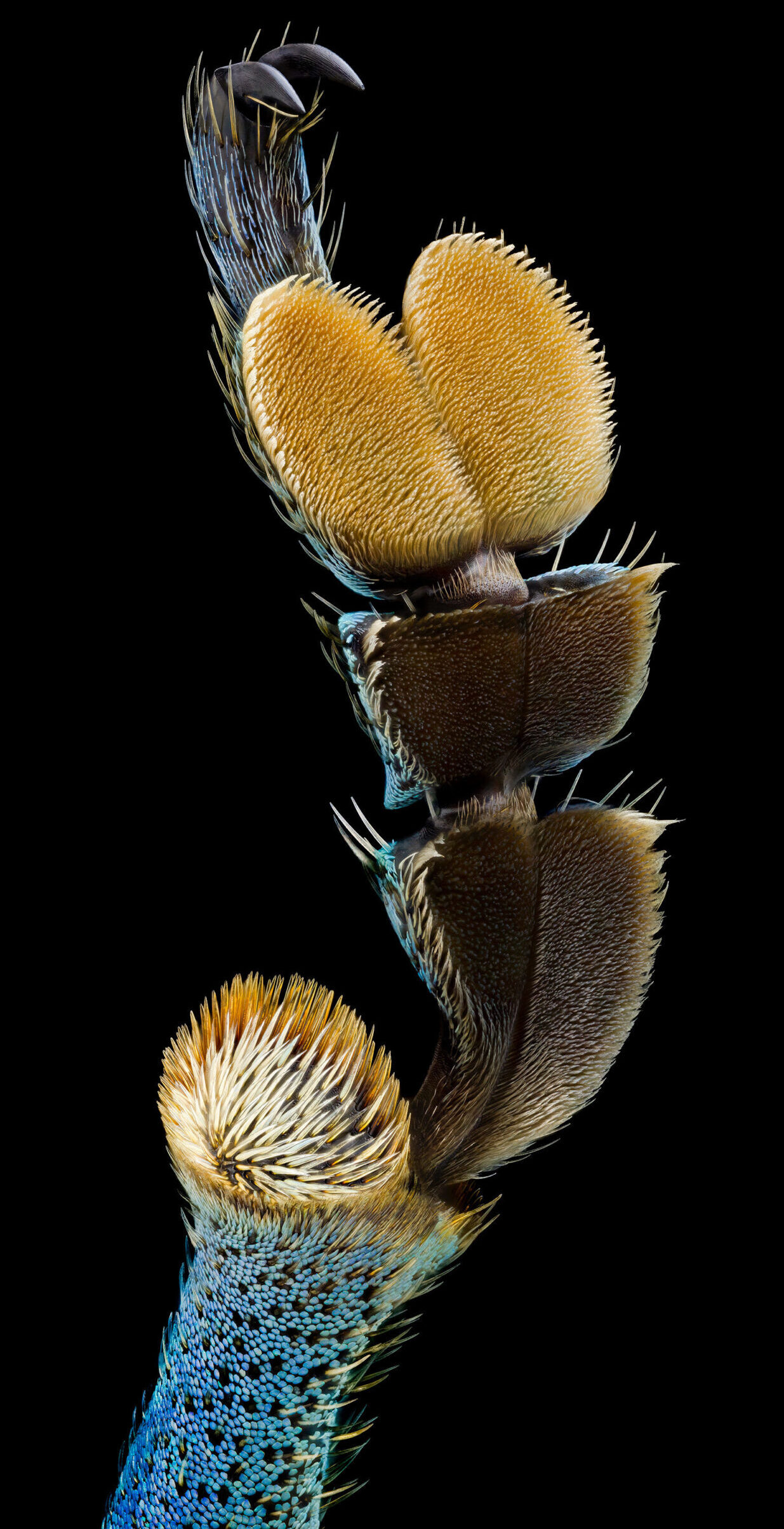
Hair on Footpad of Beetle: Nature’s Tactile Marvel
Lu-Yi Wang and Dr. Hamed Rajabi, passionate researchers in the field of microscopy, invite us to explore the microscopic wonders found on the footpad of a beetle. Through their exploration, they unravel the hidden beauty and remarkable adaptations of these tiny structures. Under the microscope, the hairs on the footpad of a beetle come alive with intricate details and fascinating shapes. These microscopic structures allow the beetle to adhere to various surfaces and navigate its environment with remarkable dexterity. This exploration celebrates the marvels of nature’s engineering and reminds us of the incredible adaptations found within even the tiniest organisms.
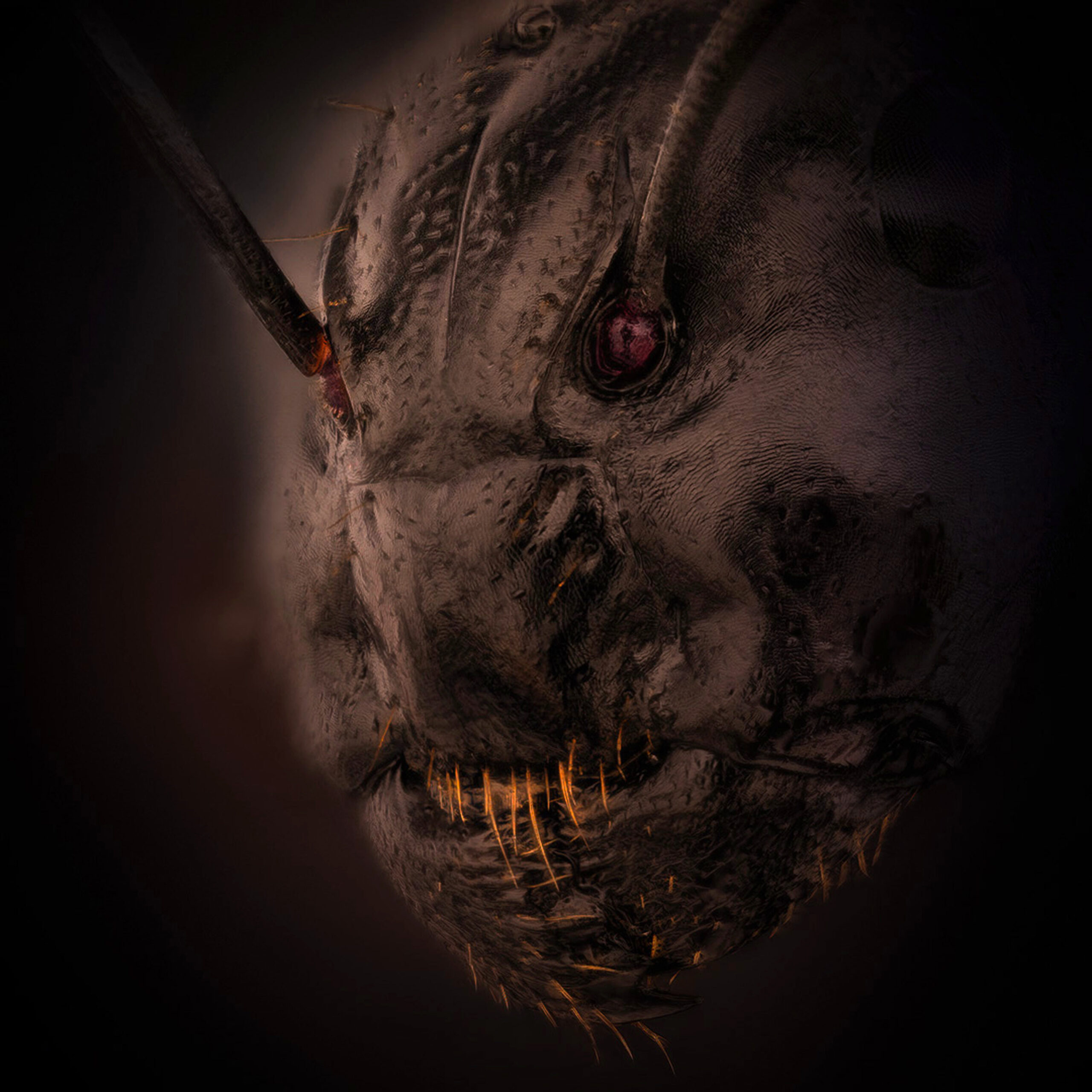
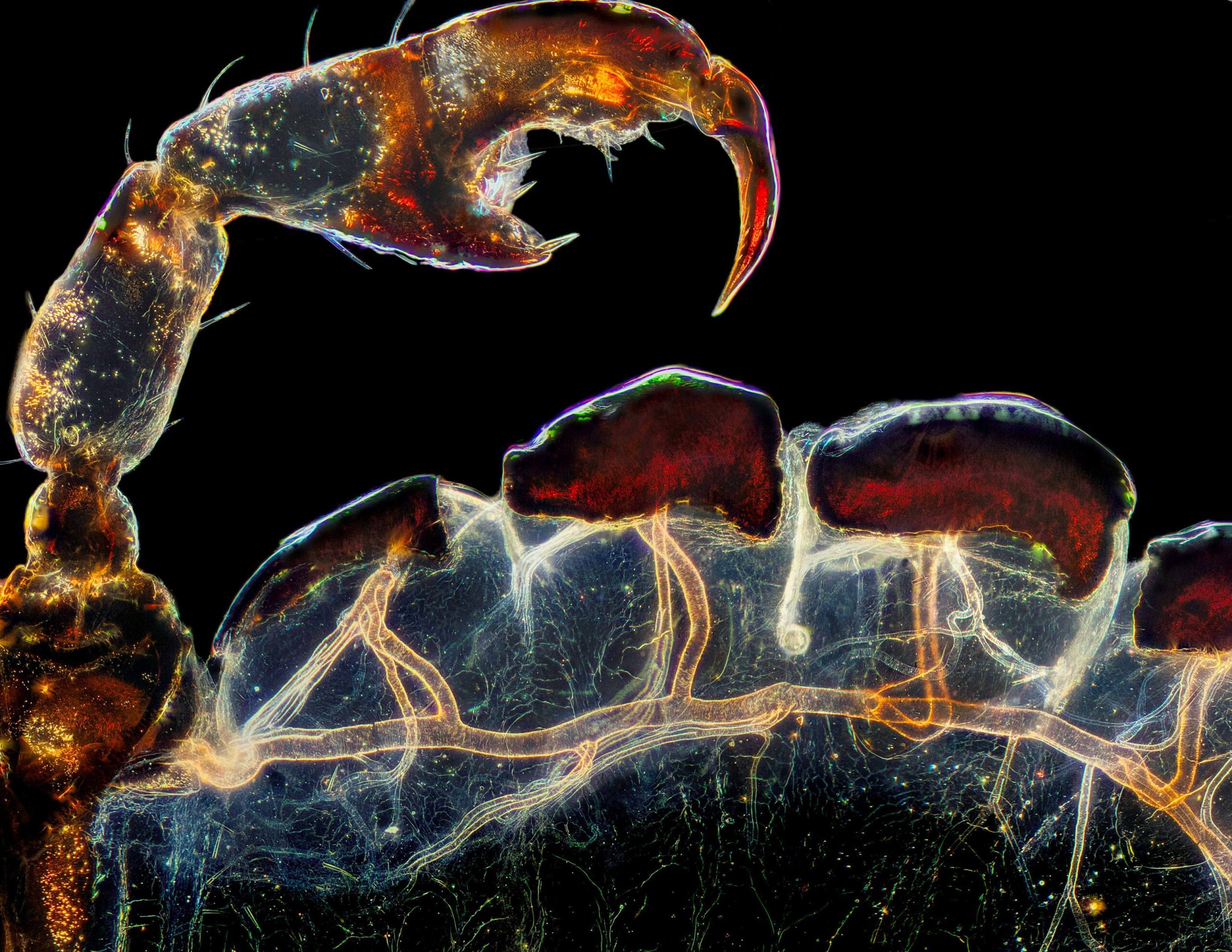
Ant: The Tiny World of Social Insects
Dr. Eugenijus Kavaliauskas, a dedicated researcher in the field of microscopy, invites us to explore the hidden world of ants. Through his lens, Kavaliauskas uncovers the intricate structures and behaviors that make these tiny creatures a fascinating subject of study. Under the microscope, ants reveal a bustling society of complex communication, organized colonies, and astonishing teamwork. Each individual ant, with its delicate exoskeleton and intricate sensory appendages, plays a crucial role in the collective success of the colony. This microscopic exploration sheds light on the remarkable adaptations and social dynamics that define the lives of ants, showcasing the beauty and complexity that exist within their miniature world.
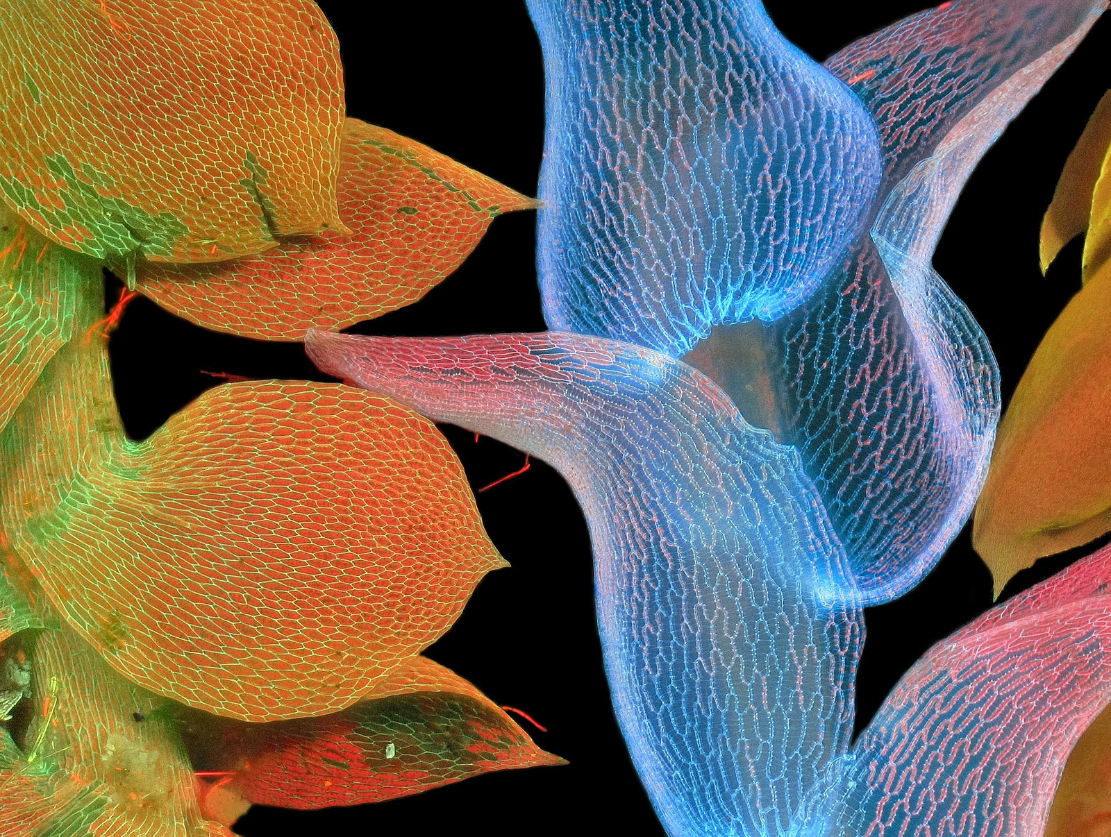
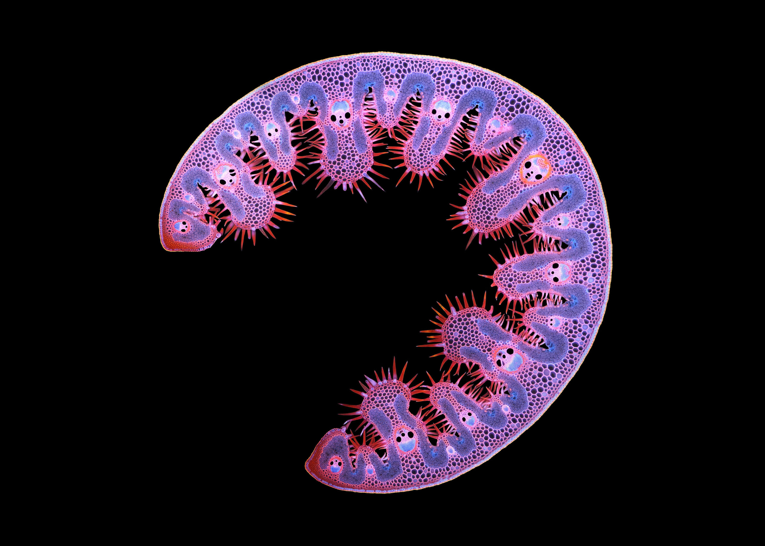
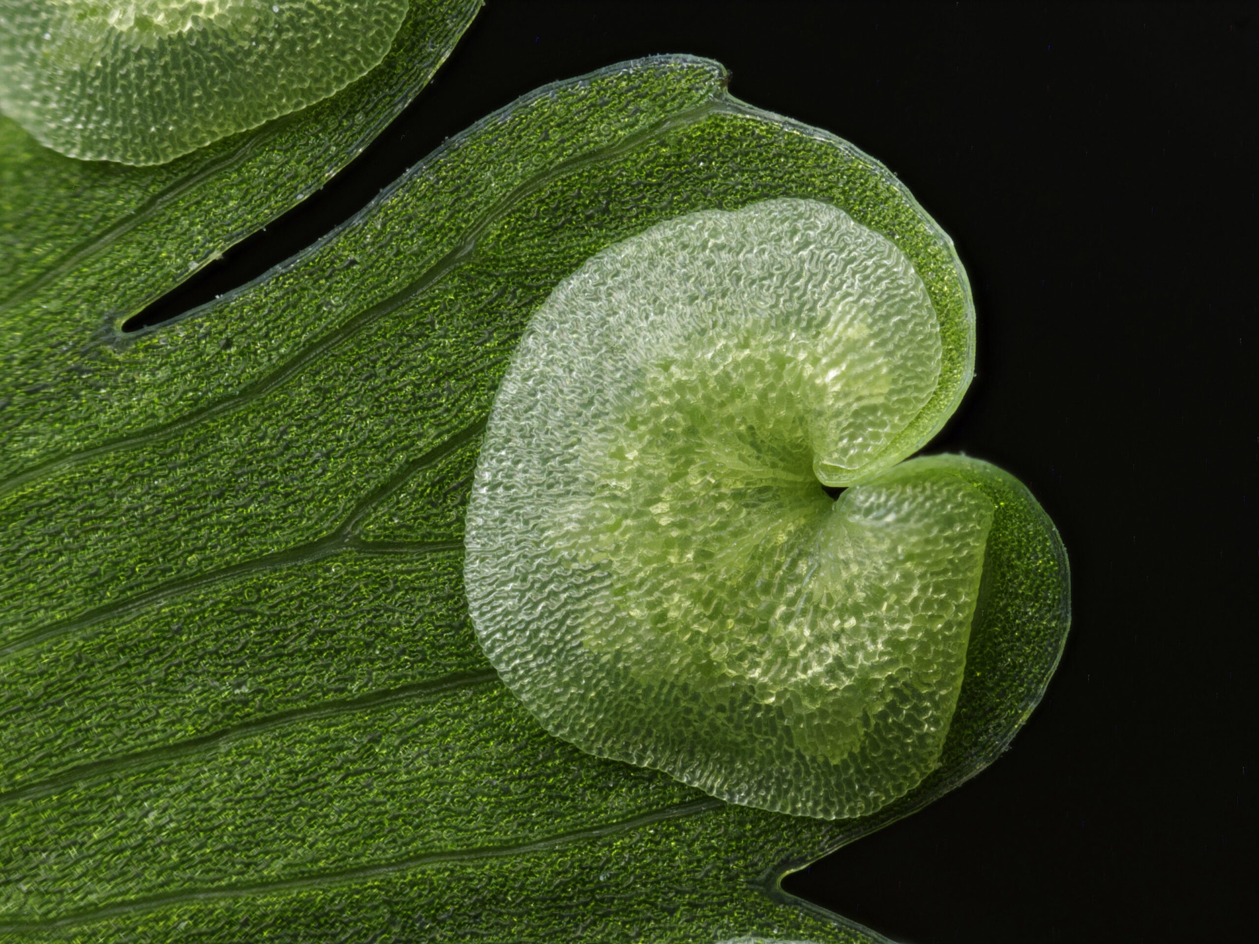
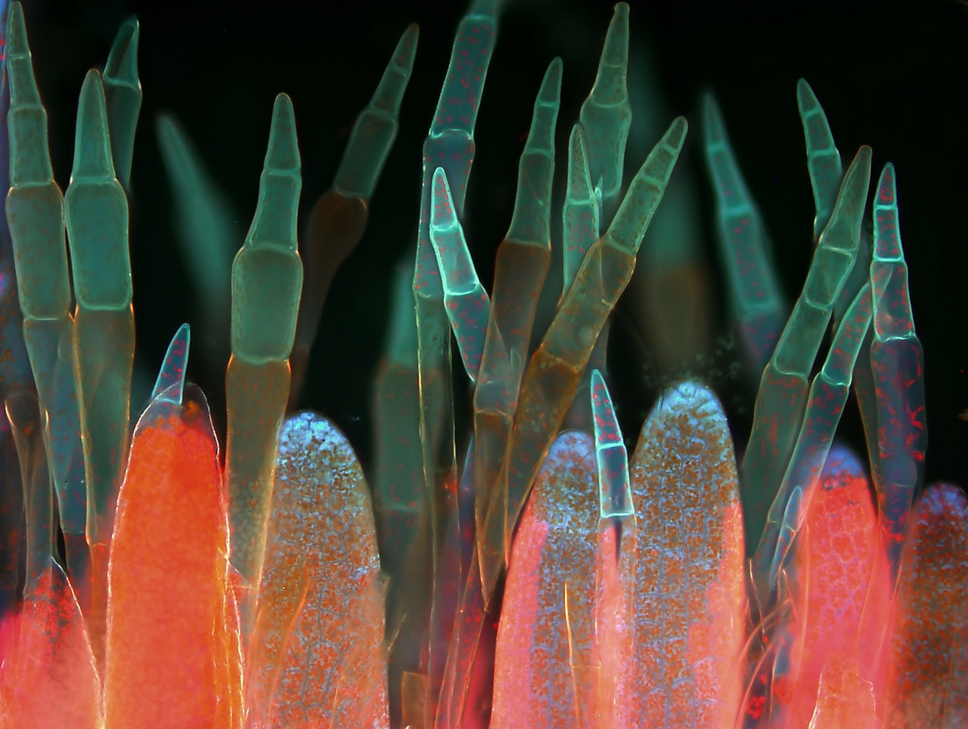
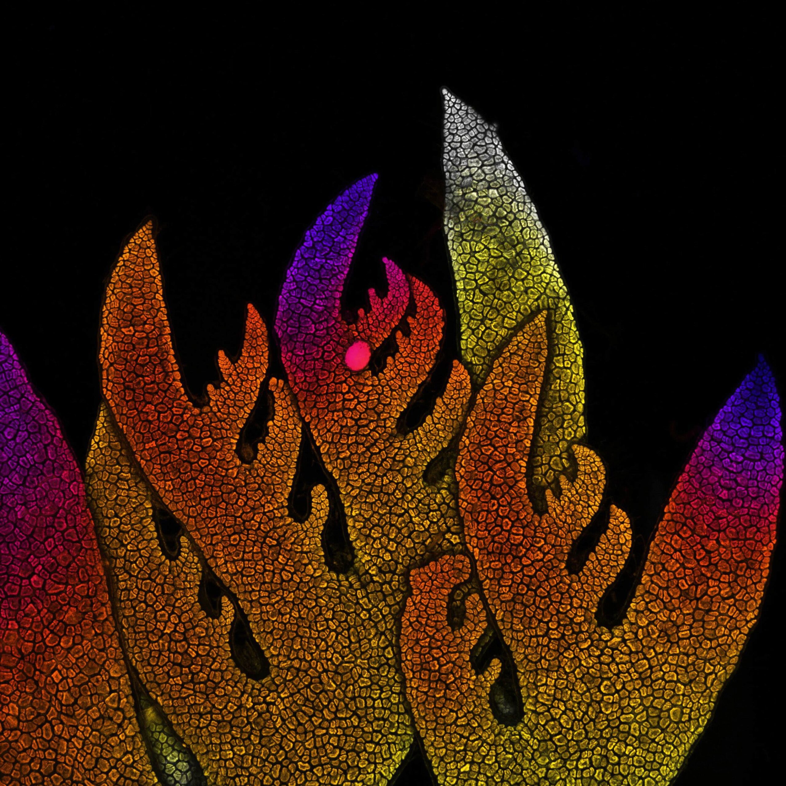
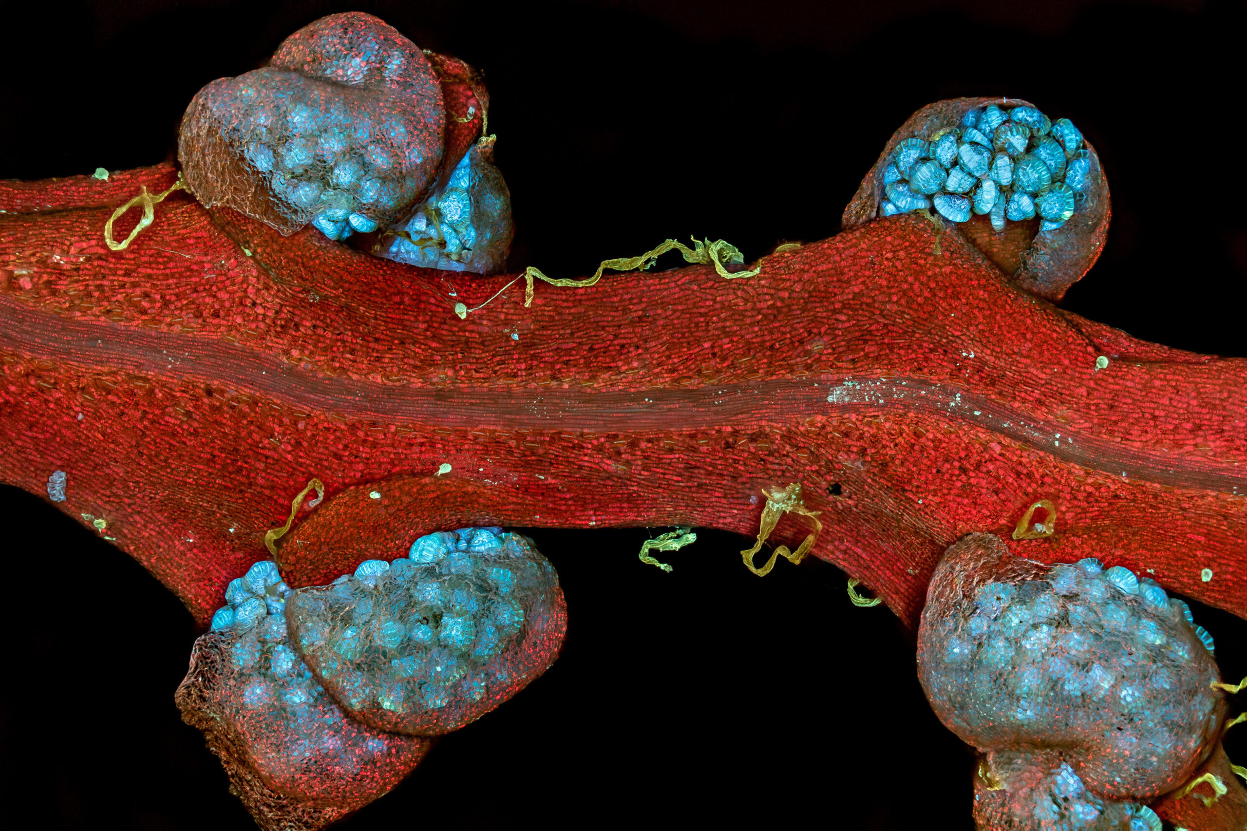
Fern Sporangium: Nature’s Reproductive Wonder
Ou Zhilei, a visionary in the world of microscopy, focuses their lens on the microscopic wonders of fern sporangia. Through their exploration, Zhilei unveils the intricate structures and reproductive mechanisms that define these ancient plants. Under the microscope, fern sporangia appear as tiny capsules of life, each housing spores that carry the potential for new growth. Delicate membranes, sporangial walls, and intricate arrangements of reproductive cells come into focus, revealing the fascinating processes that enable ferns to propagate. This exploration celebrates the resilience and elegance of nature’s reproductive mechanisms, reminding us of the intricate beauty that lies within even the most primitive of plants.
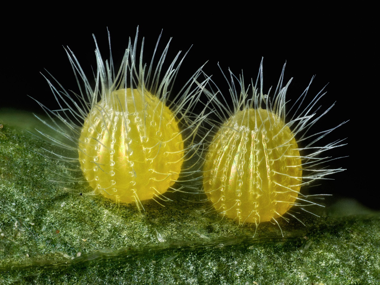
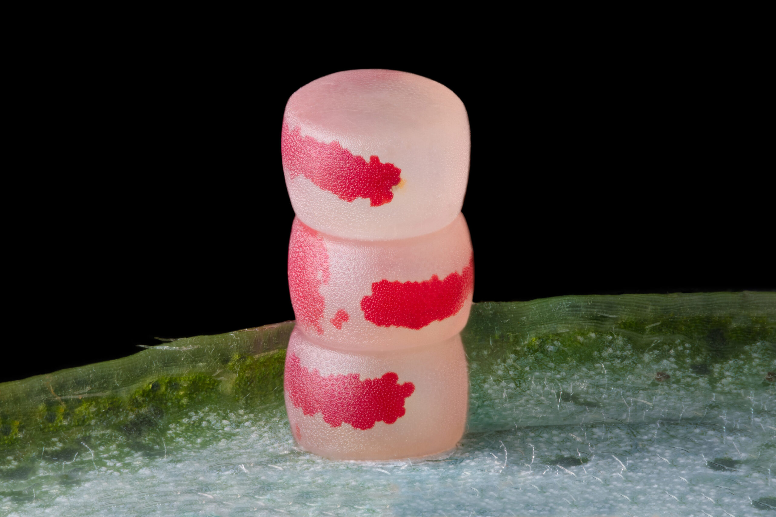
Butterfly Eggs: Nature’s Precious Beginnings
Ye Fei Zhang, a passionate microscopy enthusiast, directs our attention to the microscopic wonders found within butterfly eggs. Through their lens, Zhang unravels the hidden beauty and delicate structures that house the potential for new life. Under the microscope, butterfly eggs become captivating orbs of promise and wonder. Intricate patterns, delicate textures, and protective layers come into view, showcasing the adaptations that ensure the survival and development of future butterflies. This exploration celebrates the transformative journey of these tiny eggs, from the beginnings of life to the emergence of majestic-winged creatures.
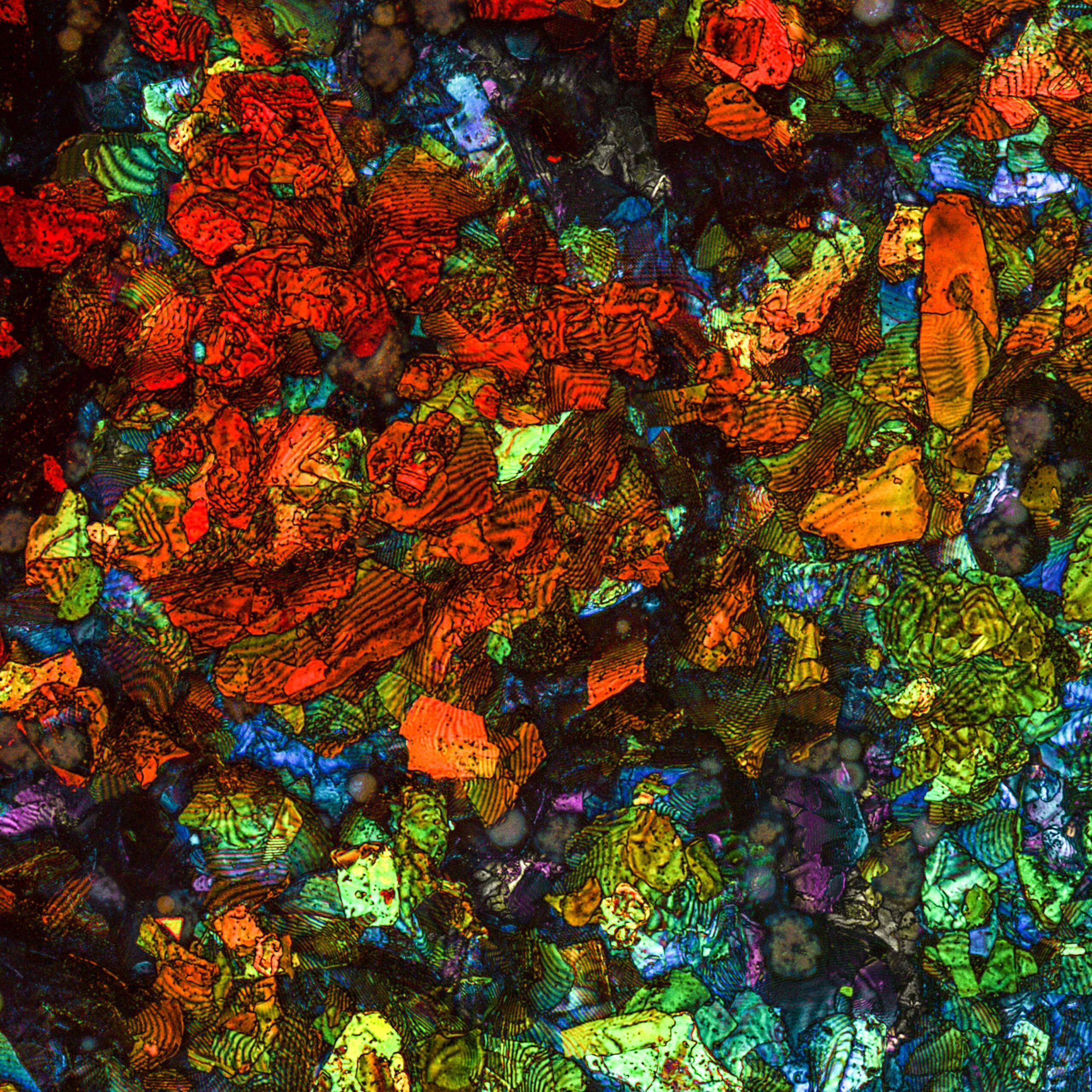
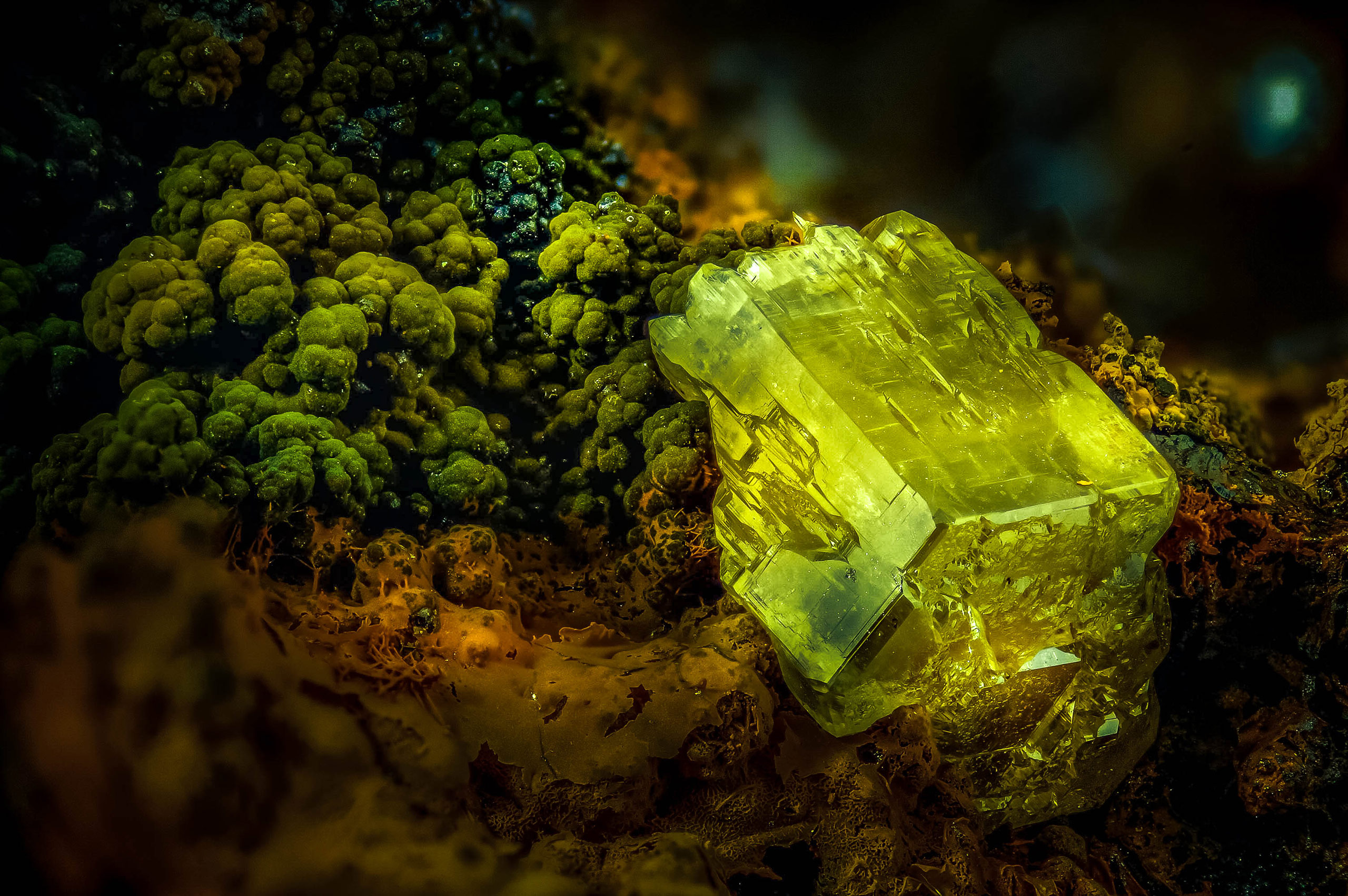
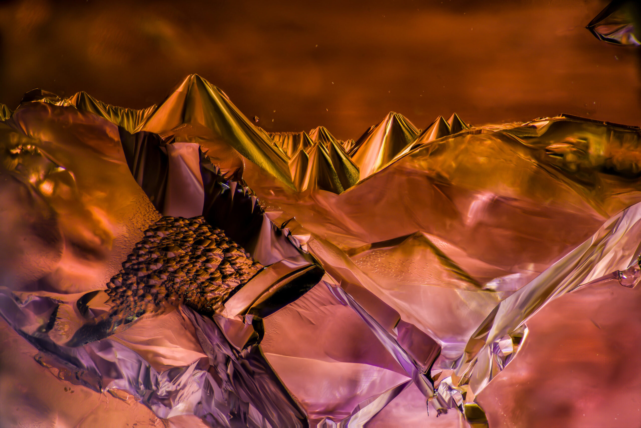
Mineral: Crystalline Marvels
Dr. Emilio Carabajal Márquez, a renowned microscopy expert, delves into the microscopic realm of minerals. Through their lens, Márquez reveals the mesmerizing beauty and intricate structures that lie within these geological treasures. Under the microscope, minerals become vibrant and captivating landscapes of crystalline formations. From the sharp edges of quartz to the lustrous hues of gemstones, each mineral reveals a unique pattern of atoms and molecules that shape its crystalline structure. This exploration invites us to appreciate the diverse array of minerals that adorn our planet, reminding us of the geological marvels hidden beneath the surface.
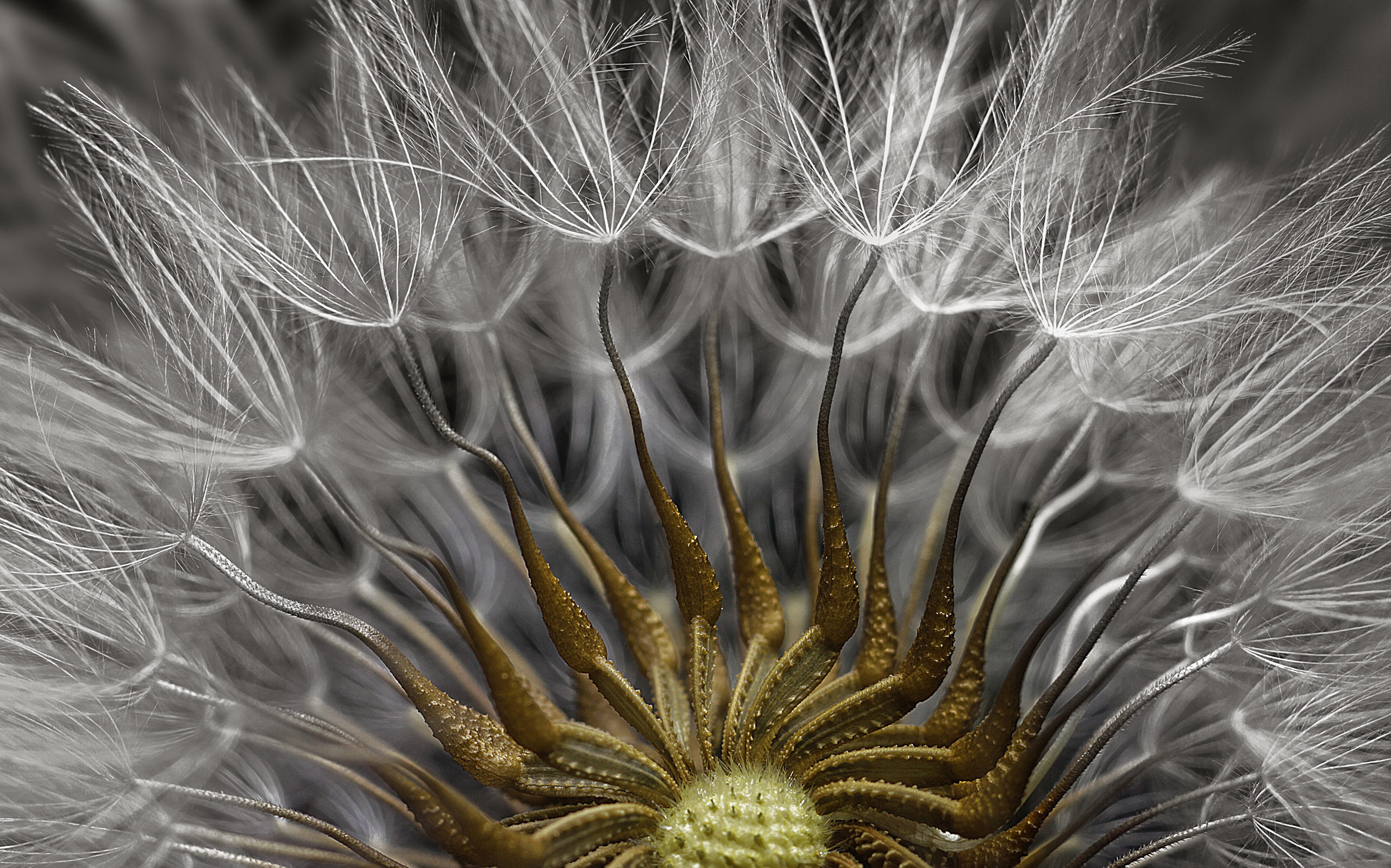
Flowering Plant Seed Head: Nature’s Fertile Abundance
Dr. Havi Sarfaty, a visionary in the world of microscopy, focuses their lens on the microscopic wonders of a flowering plant seed head. Through their exploration, Sarfaty unveils the intricate structures and reproductive potential that reside within these botanical marvels. Under the microscope, the seed head of a flowering plant comes alive with an abundance of tiny structures. Delicate seeds, protective coverings, and intricate arrangements showcase the evolutionary adaptations that enable these plants to disperse and propagate. This exploration celebrates the fertile beauty of flowering plants, reminding us of the intricate processes and remarkable diversity found within the plant kingdom.
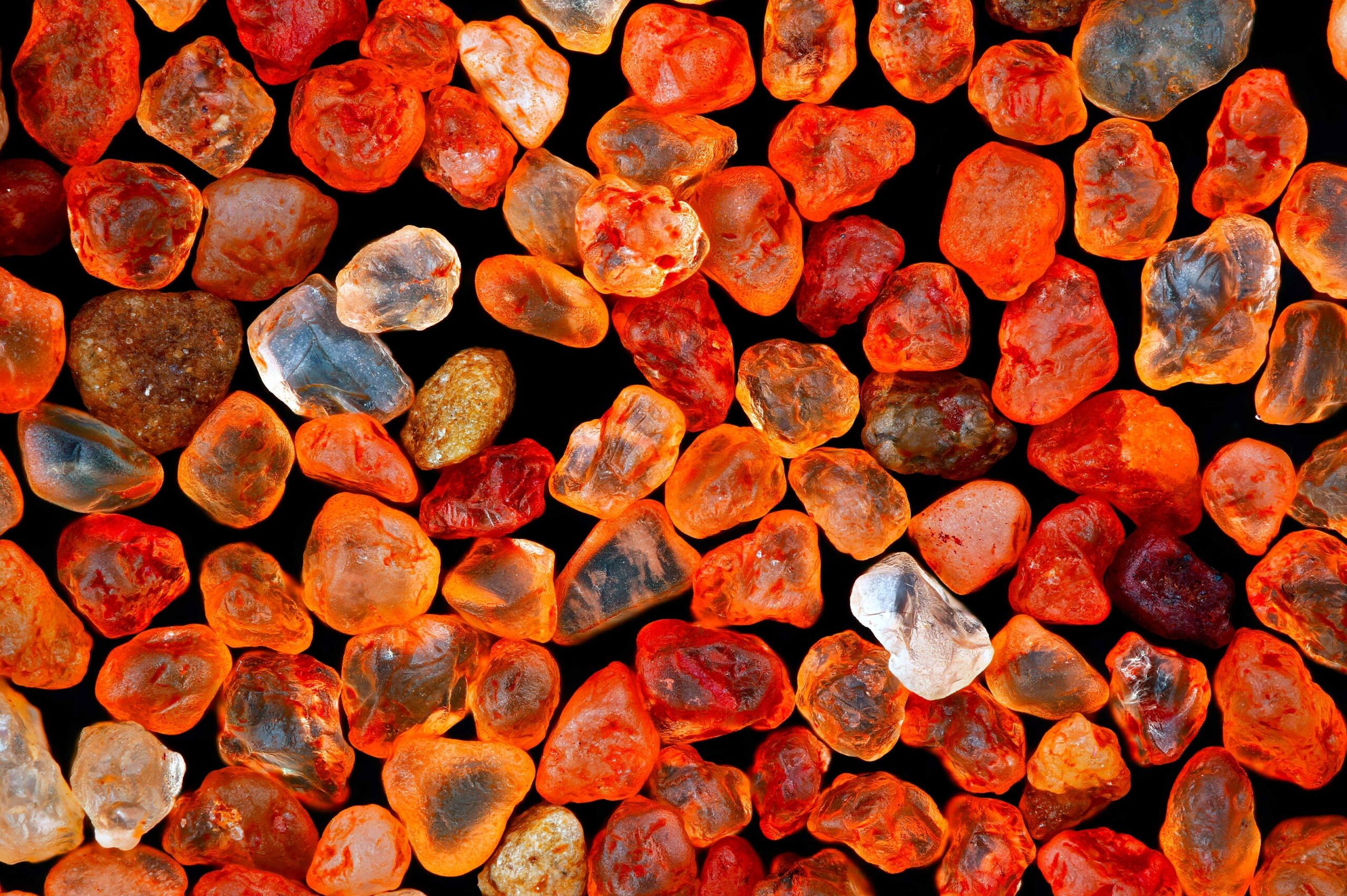
Desert Sand: Nature’s Golden Grains
Xinpei Zhang, a talented microscopy artist, takes us on a captivating journey into the microscopic world of desert sand. Through their lens, Zhang unveils the hidden beauty and intricate structures that lie within these tiny grains. Under the microscope, desert sand transforms into a mesmerizing landscape of golden hues and intricate textures. Each grain, weathered by the elements and shaped by the forces of nature, tells a unique story. From the delicate shapes of individual grains to the intricate patterns they create when gathered together, this exploration invites us to marvel at the wonders of the desert, reminding us of the remarkable diversity that exists even in the tiniest particles.
Asian Hornet: The Majesty of Nature’s Predators
Pierre Anquet, an avid explorer of the natural world, directs our attention to the microscopic wonders found within the Asian hornet. Through their lens, Anquet unravels the hidden beauty and formidable adaptations of these remarkable insects. Under the microscope, the Asian hornet reveals a world of intricate exoskeletons, delicate sensory appendages, and fearsome stingers. Each microscopic detail showcases the precision and efficiency with which these predators navigate their environment and hunt their prey. This exploration celebrates the awe-inspiring power and majesty of nature’s hunters, inviting us to appreciate the delicate balance of ecosystems and the intricate beauty of their inhabitants.
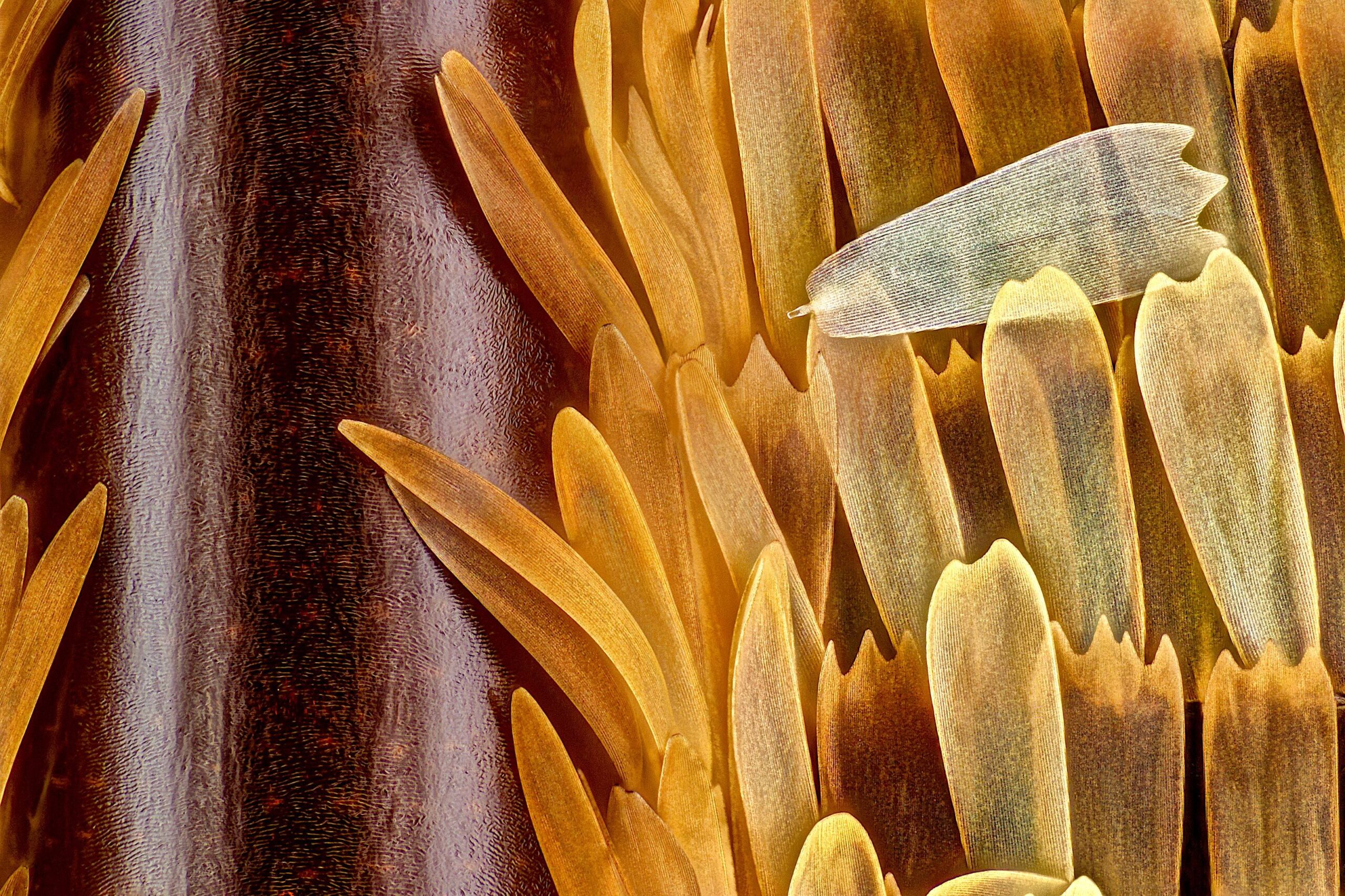
Moth Wing Scales: A Symphony of Color
Marco Jongsma, a skilled microscopy enthusiast, focuses their lens on the microscopic wonders of moth wing scales. Through their exploration, Jongsma unveils the enchanting beauty and vibrant hues that adorn these delicate structures. Under the microscope, moth wing scales become a dazzling display of colors and intricate patterns. Each scale reflects light in a unique way, creating a visual symphony that captivates the eye. This exploration celebrates the intricate beauty and remarkable adaptations found within the wings of moths, reminding us of the artistic wonders that exist within the natural world.
Red Spider Mite: Microscopic Intruders
As we dive into the microscopic world, we encounter the red spider mite—a tiny intruder that can wreak havoc on plants. Under the microscope, these minuscule pests reveal their intricate bodies, with delicate legs, exoskeletons, and mouthparts designed for survival and reproduction. This exploration serves as a reminder of the delicate balance between species and the need for harmony in our ecosystems.
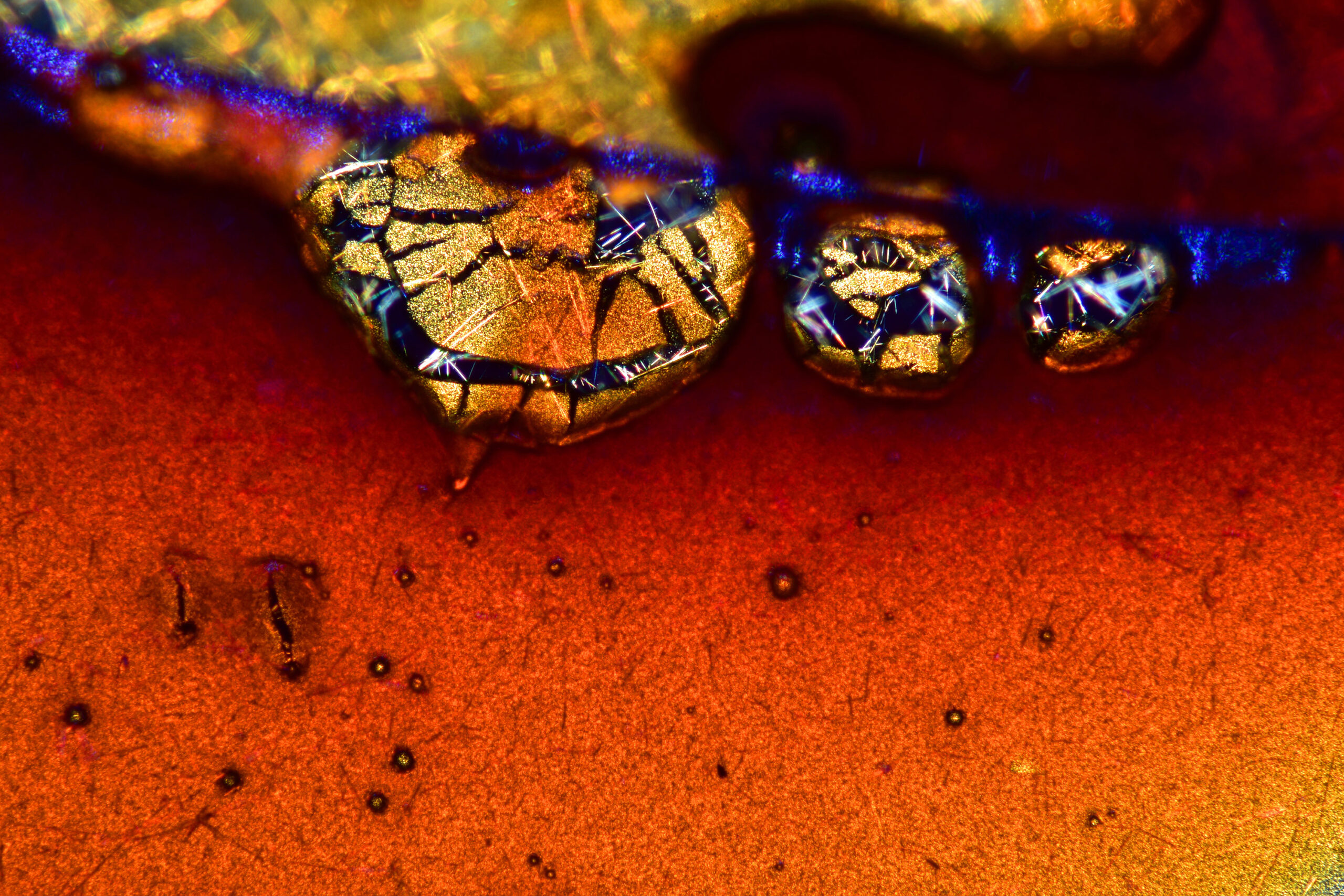
Quartz/Amethyst: Crystaline Treasures
Nick Prince, a renowned expert in the world of microscopy, invites us to explore the breathtaking beauty of quartz and amethyst crystals. Through his lens, Prince unveils the mesmerizing structures and vibrant hues that define these geological marvels. Under the microscope, quartz and amethyst crystals reveal a world of intricate formations and vibrant colors. Delicate facets, internal patterns, and the interplay of light and crystal structure create a visual spectacle that captures the imagination. This exploration celebrates the natural beauty and intricate complexity of quartz and amethyst, reminding us of the awe-inspiring wonders that lie beneath the Earth’s surface.
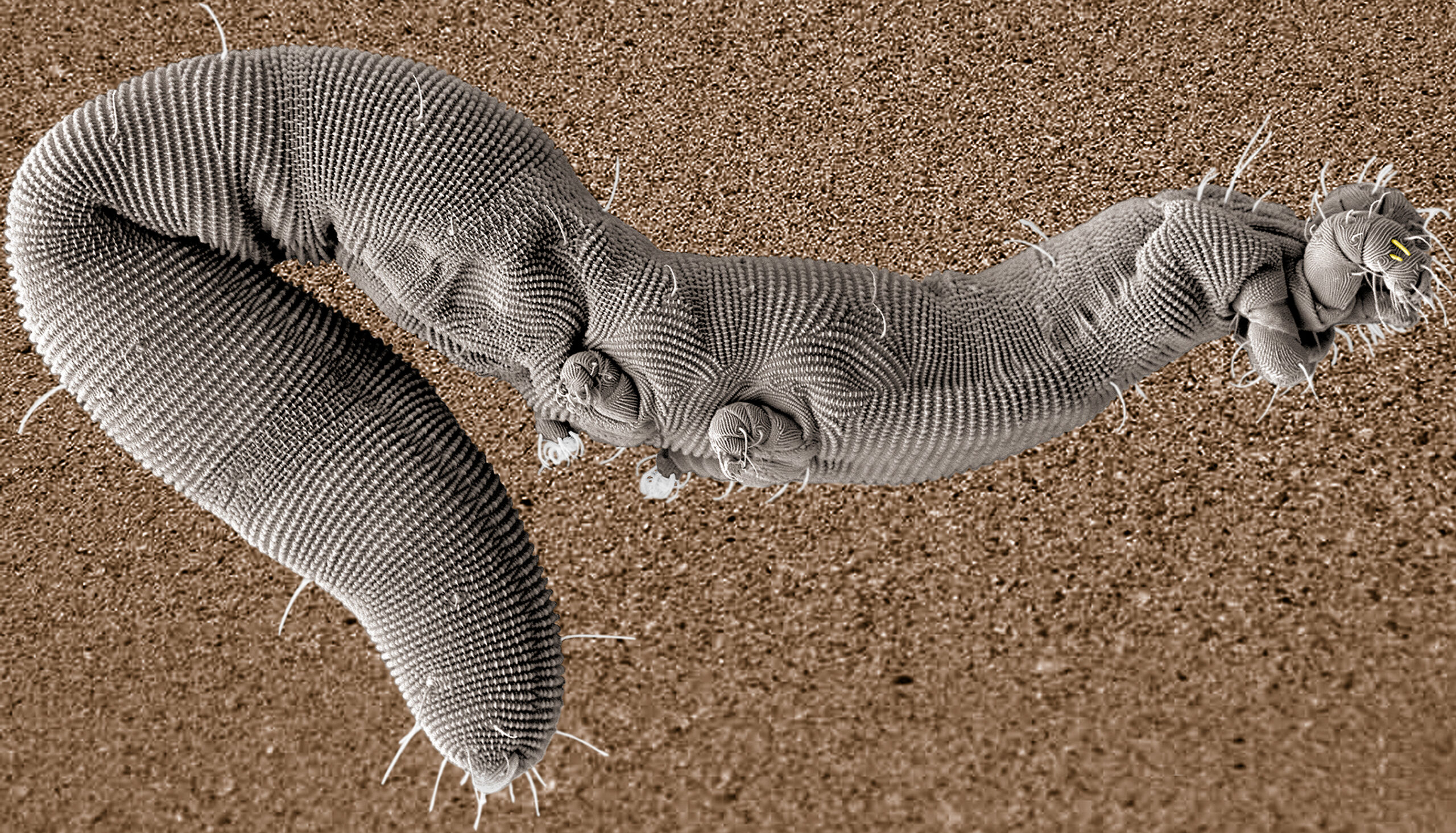
Parasite Roundworm: Unveiling Nature’s Intricate Parasitic Life
Massimo Brizzi, a dedicated researcher in the field of microscopy, directs our attention to the microscopic world of parasite roundworms. Through his lens, Brizzi unravels the hidden beauty and complex life cycles of these tiny organisms. Under the microscope, parasite roundworms reveal their intricate structures and adaptations for survival. From their specialized mouthparts to their segmented bodies, each detail showcases the remarkable strategies these organisms employ to exploit their hosts. This exploration sheds light on the delicate balance between parasites and their hosts, reminding us of the interconnectedness of all living beings.
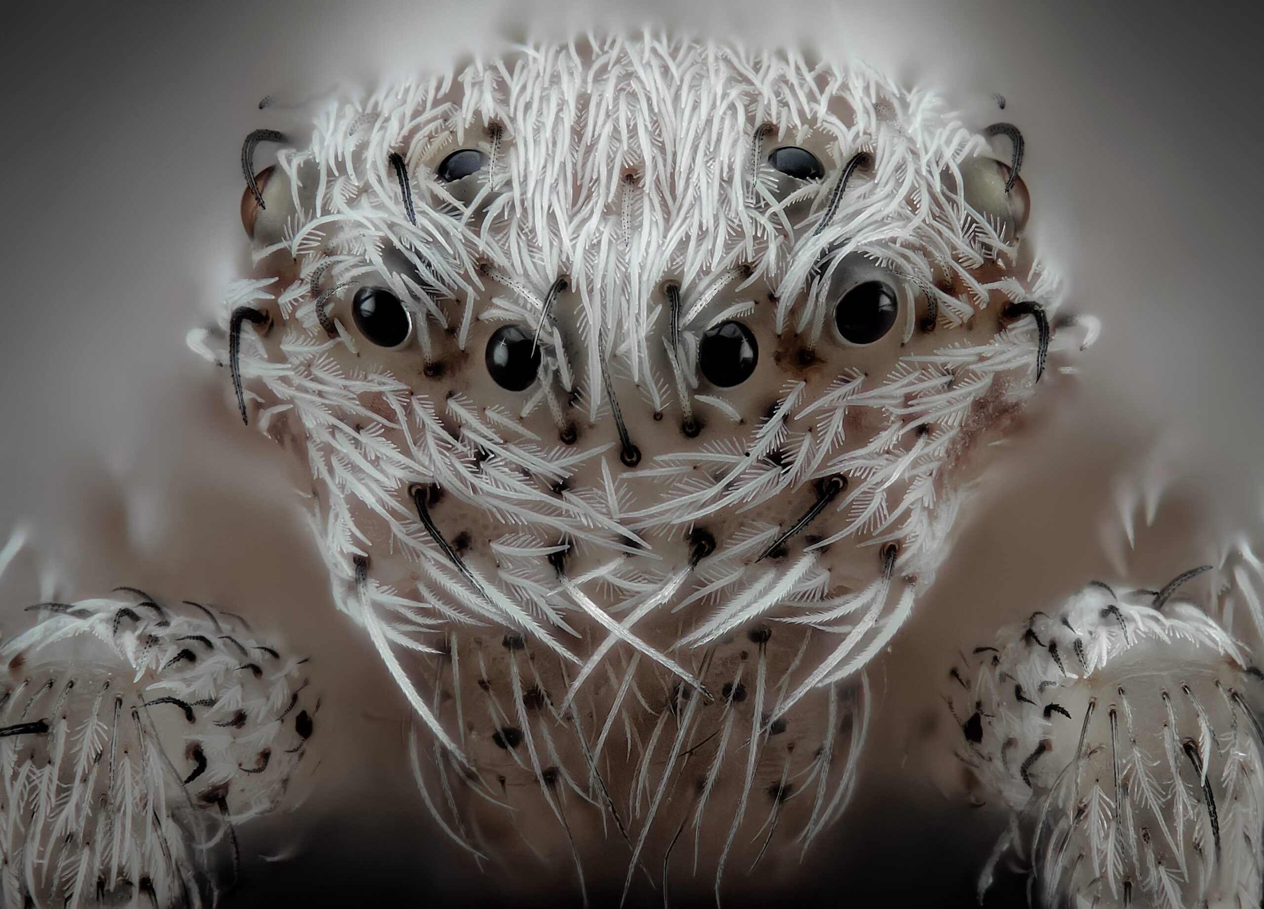
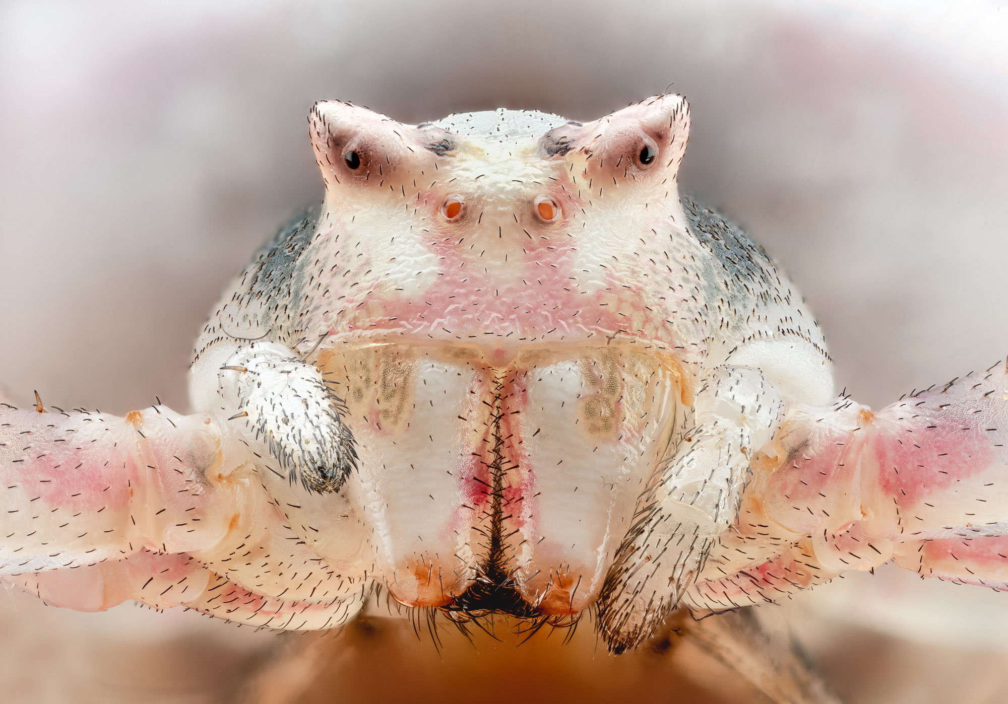
White Hairy Spider: The Elegance of Arachnid Adaptations
Javier Rupérez, a passionate microscopy enthusiast, focuses his lens on the microscopic wonders of a white hairy spider. Through his exploration, Rupérez unveils the delicate structures and intricate adaptations that define this remarkable arachnid. Under the microscope, the white hairy spider comes to life with a stunning array of fine hairs and sensory appendages. Each hair serves a purpose, allowing the spider to sense its environment and capture prey with precision. This exploration celebrates the elegance and ingenuity of arachnid adaptations, showcasing the beauty that lies within even the smallest of details.
40 Million Year Old Gnat: A Glimpse into Prehistoric Life
Levon Biss, a visionary in the world of microscopy, takes us on a journey back in time with a microscopic exploration of a 40-million-year-old gnat. Through his lens, Biss unravels the intricate structures and fossilized remains that tell the story of this ancient insect. Under the microscope, the 40-million-year-old gnat reveals preserved exoskeletons and delicate appendages. This glimpse into the past provides valuable insights into the evolution and diversity of insect life throughout history. This exploration reminds us of the vastness of geological time and the wonders that can be discovered through the lens of microscopy.
Louse: Uncovering the World of Tiny Parasites
Frank Reiser, an esteemed microscopy expert, directs our attention to the microscopic wonders found within the world of lice. Through his lens, Reiser unravels the hidden beauty and intricate adaptations of these tiny parasites. Under the microscope, lice reveal a world of complex mouthparts, specialized claws, and segmented bodies. Each microscopic detail showcases the adaptations that enable lice to attach to their hosts and feed on their blood. This exploration sheds light on the fascinating coexistence of parasites and their hosts, underscoring the intricacies of the natural world.
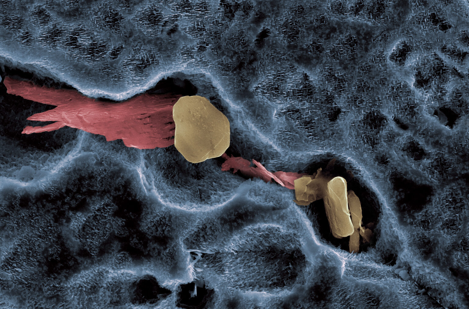
Dissolution in Nickel Superalloy: Transformations in Metal
As we delve into the microscopic world, we encounter the dissolution of nickel superalloy—an intriguing process that showcases transformations in metal. Under the microscope, we witness the intricate patterns and structural changes that occur when the superalloy comes into contact with various substances.
Moss: A Microscopic Tapestry of Nature
Magdalena Turzanska, a talented microscopy artist, invites us to peer into the enchanting world of moss. Through her lens, Turzanska unravels the intricate structures and vibrant colors that make up this miniature ecosystem. Under the microscope, moss unveils a tapestry of delicate filaments, vibrant spore capsules, and lush green structures. Each microscopic detail showcases the resilience and adaptability of these ancient plants. This exploration celebrates the beauty of moss, reminding us of the intricate wonders that exist in even the smallest corners of the natural world.
Caterpillar: The Marvels of Metamorphosis
Dean Lerman, a passionate microscopy enthusiast, focuses his lens on the microscopic wonders of a caterpillar. Through his exploration, Lerman unveils the hidden beauty and intricate structures that define the early stages of this remarkable insect’s life. Under the microscope, the caterpillar reveals its segmented body, tiny bristles, and intricate mouthparts. These microscopic details highlight the remarkable adaptations that enable caterpillars to feed and grow rapidly as they prepare for their transformative journey into adulthood. This exploration showcases the wonders of metamorphosis and the intricate beauty that lies within the life cycles of insects.
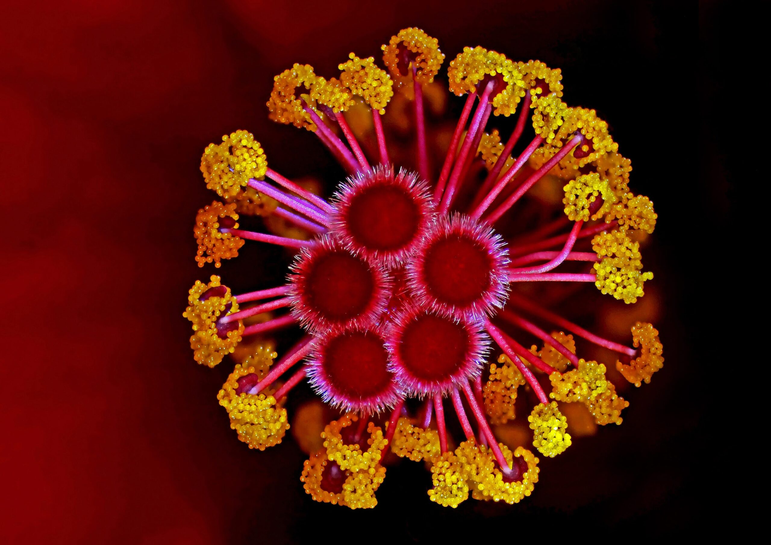
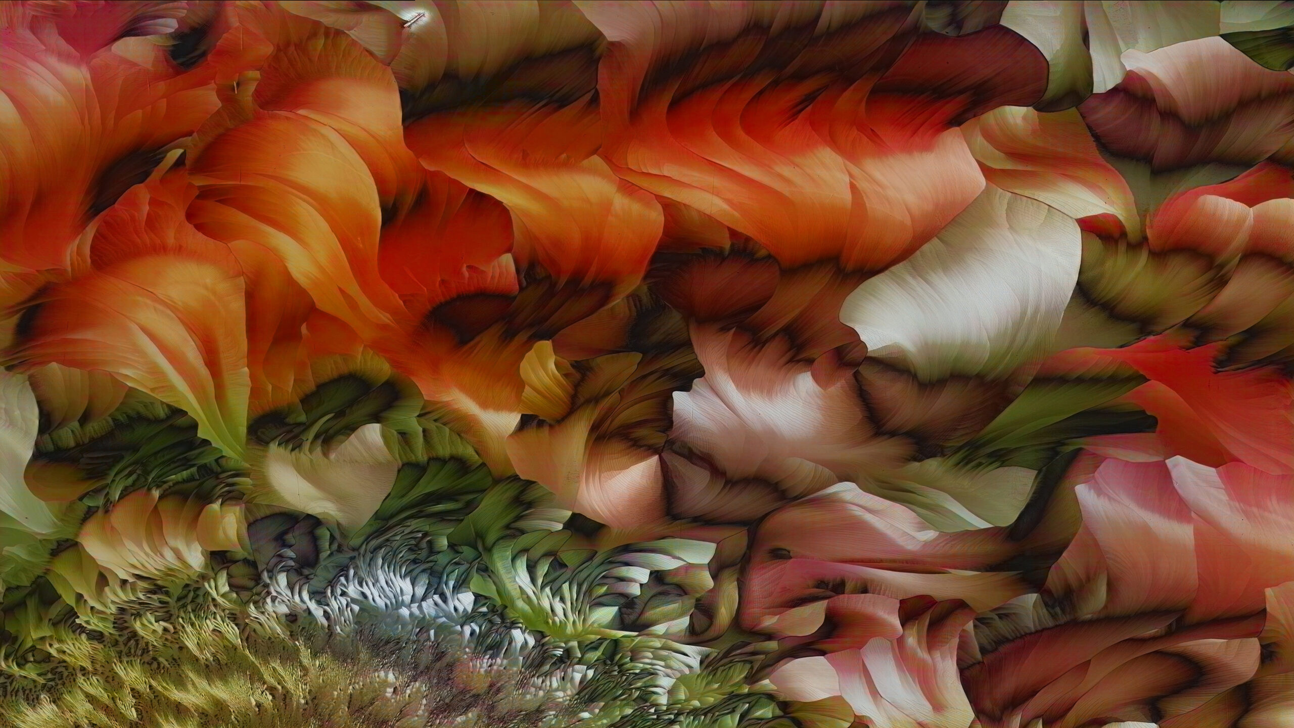
Hibiscus: A Floral Symphony in Microscopic Detail
Dr. Csaba László Pintér, a visionary in the world of microscopy, directs our attention to the microscopic wonders found within the petals of a hibiscus flower. Through his exploration, Pintér unveils the intricate structures and vibrant colors that adorn these botanical marvels. Under the microscope, the hibiscus flower comes alive with an array of delicate petal structures, vibrant pigments, and intricate patterns. Each microscopic detail showcases the intricacies of floral biology, from the unique arrangement of cells to the specialized structures that aid in pollination. This exploration celebrates the captivating beauty of hibiscus flowers, reminding us of the remarkable diversity and sophistication found within the plant kingdom.
Slime Mold: A Microscopic Wonder of Nature
Alison Pollack, a dedicated researcher in the field of microscopy, unveils the hidden beauty and peculiar behavior of slime molds. Through her lens, Pollack explores the intricate structures and vibrant colors that make these organisms both fascinating and mysterious. Under the microscope, slime molds reveal a network of delicate filaments and fruiting bodies that showcase their ability to navigate their environment and form complex patterns. This exploration sheds light on the unique characteristics of slime molds, highlighting their role in decomposition and their intriguing life cycles. It is a reminder of the diversity and resilience of life, even in the most unexpected forms.
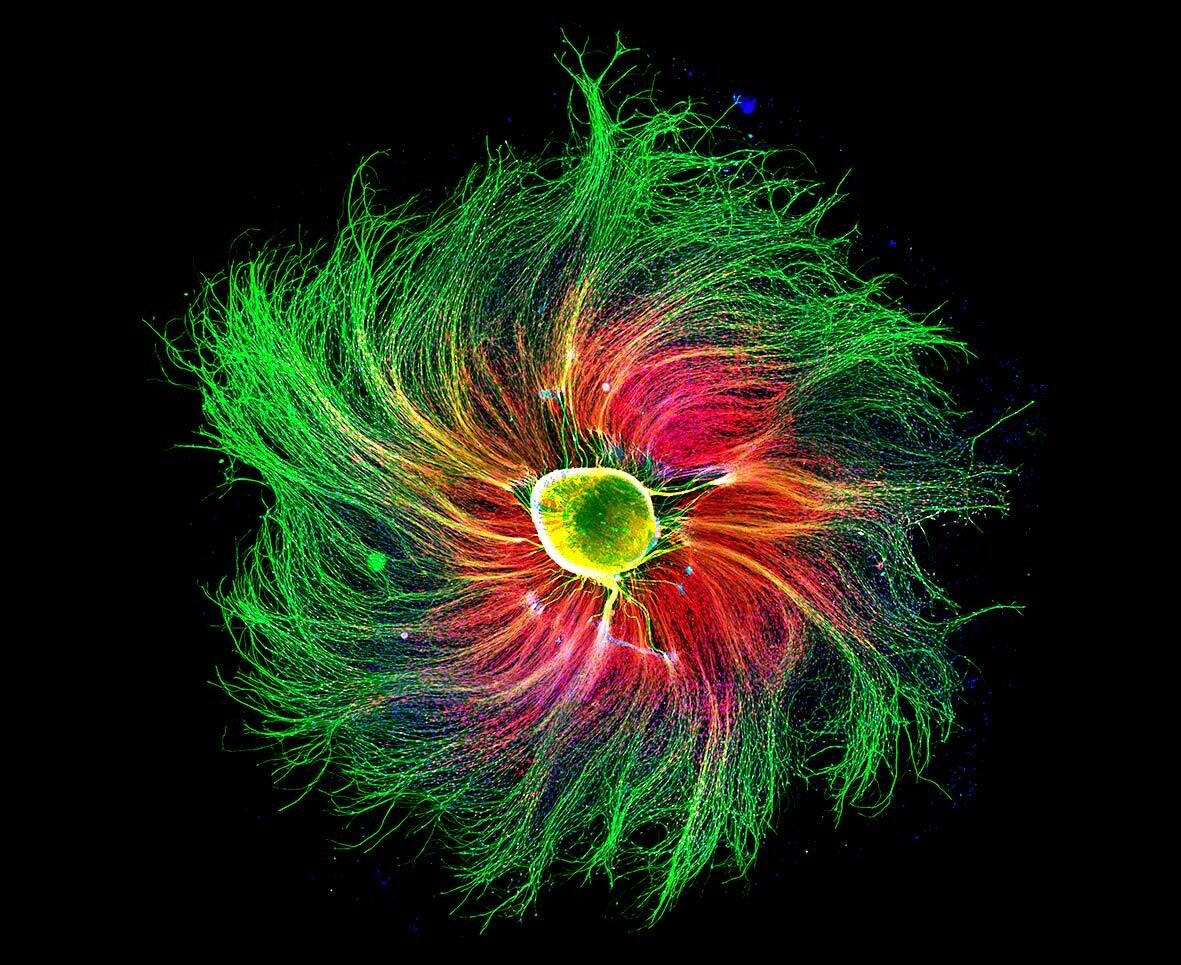
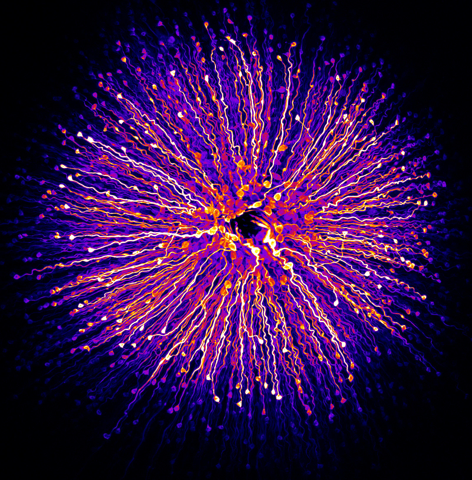
Sensory Neuron of Rat: The Intricacies of Perception
Paula Diaz, an esteemed expert in the world of microscopy, focuses her lens on the microscopic wonders of a sensory neuron in a rat. Through her exploration, Diaz unravels the intricate structures and connections that enable these neurons to transmit signals and perceive the world around them. Under the microscope, the sensory neuron reveals a complex network of dendrites, axons, and synapses. Each microscopic detail showcases the remarkable precision and complexity of the nervous system, highlighting the role of sensory neurons in perceiving and interpreting sensory information. This exploration celebrates the wonders of neuroscience and the intricacies of perception in the animal kingdom.
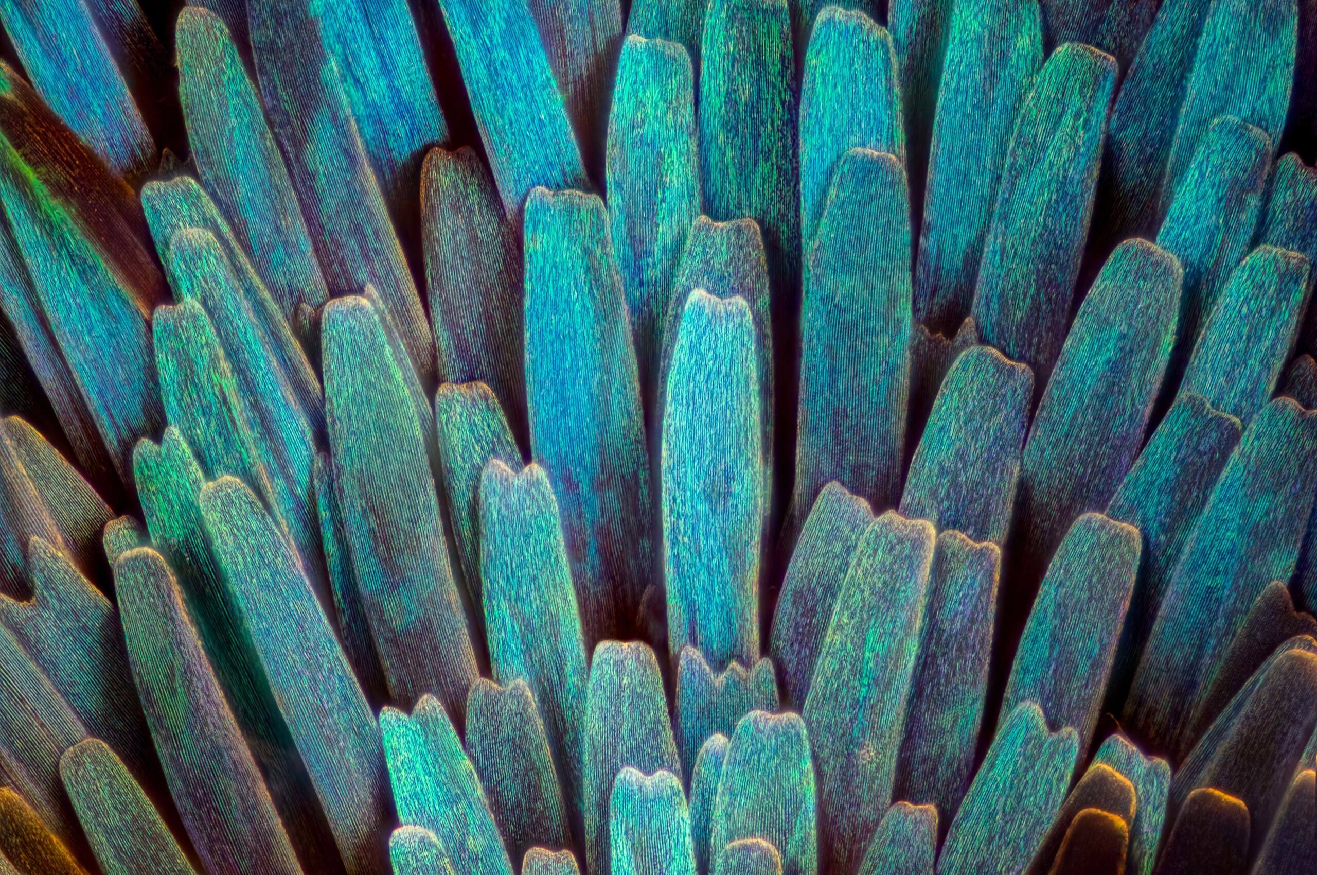
Butterfly Wing Scales: A Kaleidoscope of Color
Luciano Andres Richino, a skilled microscopy artist, takes us on a mesmerizing journey into the microscopic world of butterfly wing scales. Through his lens, Richino unveils the intricate structures and vibrant colors that make these delicate wings a visual spectacle. Under the microscope, butterfly wing scales reveal a symphony of colors, intricate patterns, and fine textures. Each scale reflects light in a unique way, creating a dazzling display that captivates the eye. This exploration celebrates the artistic marvels found within the natural world, reminding us of the incredible diversity and beauty that exist in even the tiniest of creatures.
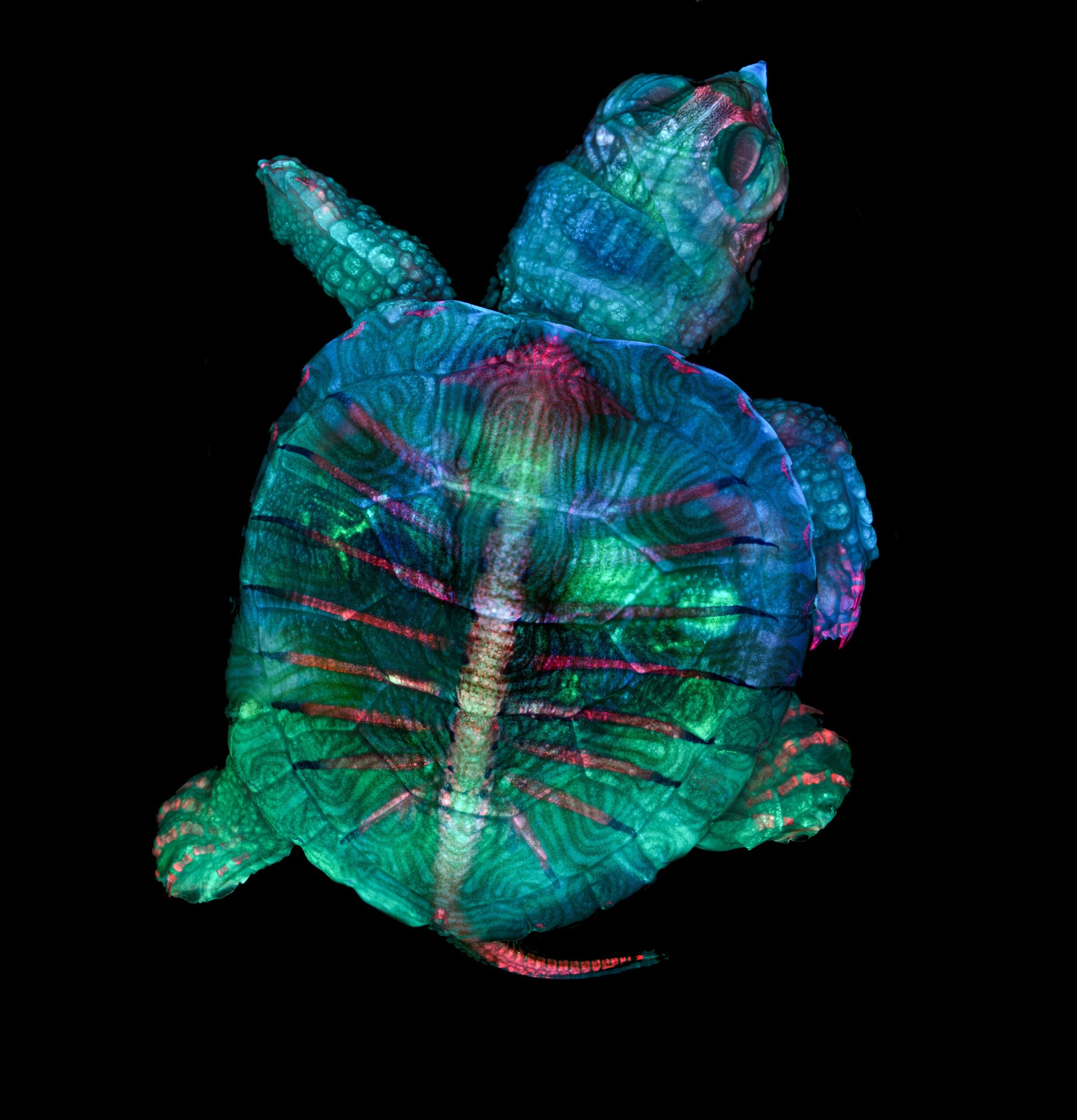
Fluorescent Turtle Embryo: Illuminating Life’s Beginnings
Teresa Zgoda and Teresa Kugler, renowned microscopy experts, shed light on the earliest stages of life through their exploration of a fluorescent turtle embryo. Through their lens, Zgoda and Kugler unveil the intricate structures and vibrant fluorescence that highlight the development of these fascinating creatures. Under the microscope, the turtle embryo reveals delicate tissues, developing organs, and intricate blood vessels, all bathed in mesmerizing fluorescent colors. This exploration provides a glimpse into the wonders of embryonic development and the remarkable processes that shape life from its earliest moments. It serves as a reminder of the intricate beauty and resilience found within the natural world.
Fumarolic Incrustations: Nature’s Geological Art
Exploring the microscopic wonders of fumarolic incrustations, we uncover the remarkable formations that result from volcanic activity. Under the microscope, these incrustations showcase intricate crystal structures, vibrant colors, and intricate patterns formed by mineral deposition. This exploration celebrates the artistic creations of nature, reminding us of the dynamic forces at work beneath the Earth’s surface.

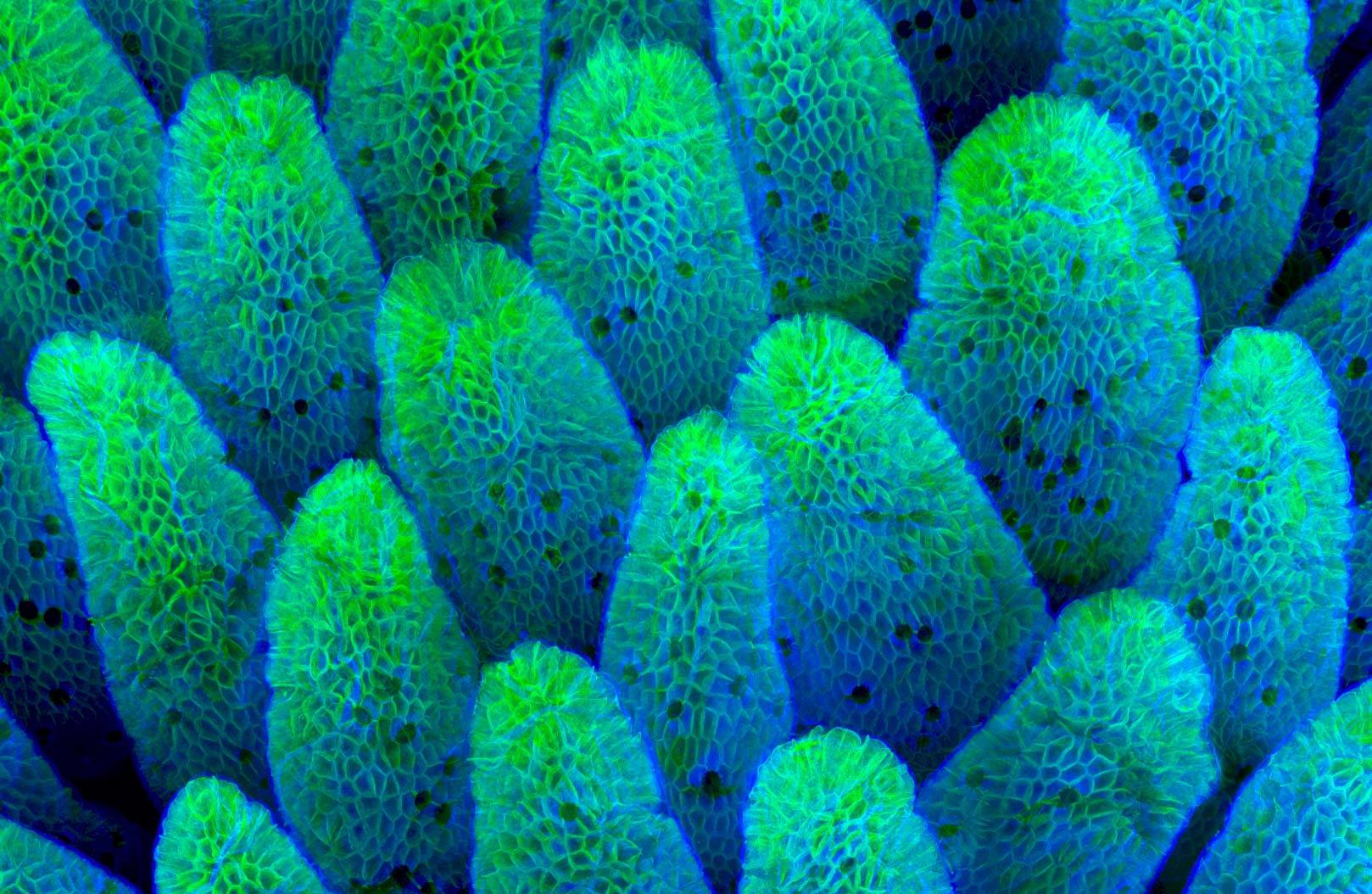
Growing Tip of a Red Algae: The Beauty of Botanical Growth
Dr. Nathanaël Prunet, a visionary in the field of microscopy, directs our attention to the growing tip of a red algae. Through his lens, Prunet unravels the intricate structures and vibrant colors that define the growth and development of this botanical marvel. Under the microscope, the growing tip of the red algae reveals delicate cells, meristematic tissues, and intricate patterns of growth. Each microscopic detail showcases the remarkable processes that allow plants to continuously adapt and thrive in their environments. This exploration celebrates the beauty of botanical growth and the intricacies of plant biology.
Tulip Bud Cross Section: A Floral Journey Unveiled
Andrei Savitsky, a passionate microscopy enthusiast, focuses his lens on the cross-section of a tulip bud. Through his exploration, Savitsky unveils the hidden beauty and intricate structures that define the early stages of a tulip’s development. Under the microscope, the tulip bud cross section reveals a tapestry of delicate petals, intricate reproductive structures, and vascular networks. Each microscopic detail showcases the intricate processes that govern the growth and flowering of plants. This exploration celebrates the beauty and complexity of plant anatomy, reminding us of the wonders that lie within the natural world.
Amino Acid Crystals: Illuminating the Building Blocks of Life
Dr. John Hart, a pioneering researcher, invites us to delve into the microscopic world of amino acid crystals. Through his lens, Hart unravels the intricate structures and vibrant colors that make up these fundamental building blocks of life. Under the microscope, amino acid crystals reveal a symphony of geometric patterns and delicate arrangements. Each crystal showcases the precise interactions between individual molecules, forming the foundation of proteins and the complex biological processes they enable. This exploration celebrates the elegance and beauty of molecular structures, reminding us of the remarkable intricacies that underlie life itself.
Moth Eggs: The Start of a Lepidopteran Journey
Ye Fei Zhang, a dedicated microscopy enthusiast, directs our attention to the microscopic wonders found within moth eggs. Through their exploration, Zhang unveils the intricate structures and hidden beauty that mark the beginning of these fascinating insects’ life cycles. Under the microscope, moth eggs showcase a variety of shapes, sizes, and textures. Each microscopic detail reveals the adaptability and survival strategies of these tiny organisms as they prepare to embark on their transformational journey into adulthood. This exploration highlights the marvels of insect development and the incredible diversity that exists within the natural world.
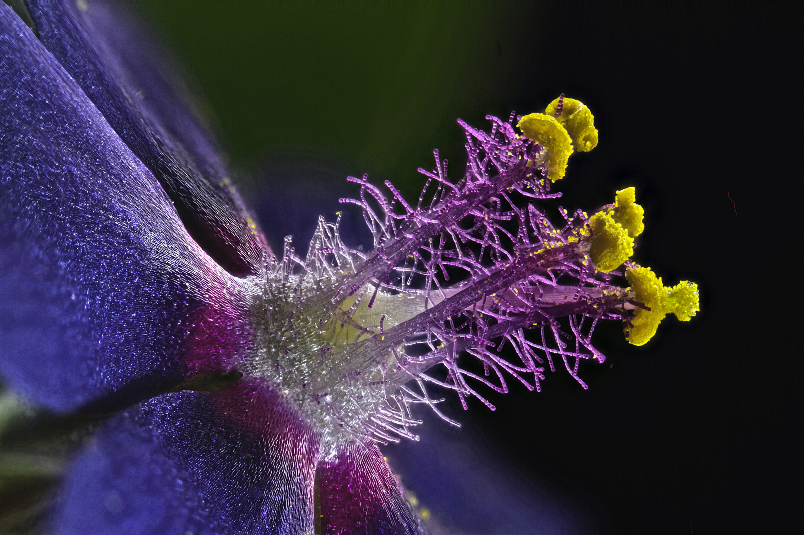
Wildflower Stamens: Nature’s Intricate Reproductive Artistry
Samuel Silberman, a talented microscopy artist, focuses his lens on the microscopic wonders found within the stamens of wildflowers. Through his exploration, Silberman unveils the intricate structures and vibrant colors that define these essential reproductive organs. Under the microscope, wildflower stamens come alive with delicate filaments, pollen grains, and intricate floral arrangements. Each microscopic detail showcases the diverse strategies employed by plants to attract pollinators and ensure their reproductive success. This exploration celebrates the intricate artistry of nature’s reproductive processes, reminding us of the interconnectedness between plants and the pollinators that rely on them.
Midge: Exploring the Microscopic Inhabitants of Water
Dr. Erick Francisco Mesén, a passionate researcher, invites us to peer into the microscopic world of a midge. Through their lens, Mesén unravels the hidden beauty and intricate structures that define the life of this tiny aquatic creature. Under the microscope, the midge reveals delicate body segments, feathery appendages, and intricate mouthparts. Each microscopic detail showcases the adaptations that enable these organisms to thrive in aquatic environments and play crucial roles in the ecological balance of freshwater ecosystems. This exploration sheds light on the marvels of aquatic biodiversity and the interconnectedness of life within watery realms.
Zebrafish: A Microscopic Glimpse into Vertebrate Development
Dr. Julien Resseguier, a visionary in the field of microscopy, focuses their lens on the microscopic wonders found within zebrafish. Through their exploration, Resseguier unravels the intricate structures and dynamic processes that define the development of these transparent and genetically versatile vertebrates. Under the microscope, zebrafish embryos reveal delicate tissues, developing organs, and intricate vascular networks. Each microscopic detail showcases the astonishing capacity of these organisms for rapid growth and regeneration, making them invaluable models for studying human development and disease. This exploration celebrates the beauty and complexity of vertebrate embryology, offering insights into the mysteries of life’s early stages.
Retina: Unveiling the Visionary World
Hanen Khabou, an accomplished microscopy artist, directs our attention to the intricate structures of the retina. Through her lens, Khabou reveals the hidden beauty and complex architecture that underlies our visual perception. Under the microscope, the retina unfolds as a delicate network of specialized cells, including photoreceptors, ganglion cells, and intricate neural connections. Each microscopic detail highlights the remarkable adaptation of this sensory organ, showcasing its role in converting light into electrical signals that allow us to see and interpret the world around us. This exploration celebrates the wonders of vision and the intricacies of the human visual system.
Human Hair: A Microcosm of Individuality
Robert Vierthaler, a passionate microscopy enthusiast, focuses his lens on the microscopic wonders found within a strand of human hair. Through his exploration, Vierthaler unravels the intricate structures and diverse characteristics that make each strand of hair a unique identifier. Under the microscope, human hair reveals a fascinating array of cuticles, medulla, and cortical cells. Each microscopic detail showcases the distinct qualities of different hair types, colors, and textures. This exploration celebrates the individuality and diversity found within human biology, reminding us that even the smallest aspects of our bodies can tell a unique story.
Plant Tendril: Nature’s Grasping Grace
Saulius Gugis, a talented microscopy artist, invites us to appreciate the intricate beauty of a plant tendril. Through his lens, Gugis unveils the hidden structures and elegant curves that allow these botanical appendages to navigate and anchor themselves in their environment. Under the microscope, plant tendrils reveal delicate cells, flexible fibers, and intricate curling mechanisms. Each microscopic detail showcases the remarkable adaptability and grace of these structures, enabling plants to climb, grasp, and reach for support. This exploration celebrates the wonders of plant morphology and the fascinating strategies employed by plants to secure their place in the natural world.
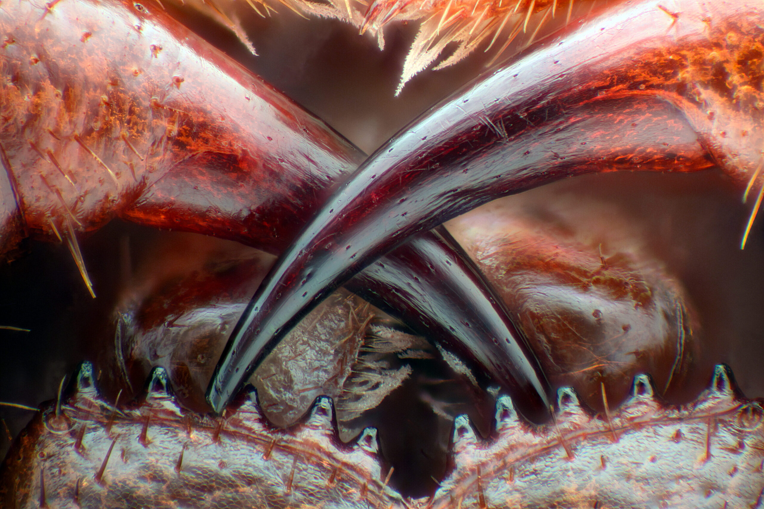
Poison Fangs of Centipede: Venomous Delicacy
Walter A. Piorkowski, a pioneering researcher in the field of microscopy, focuses his lens on the microscopic wonders found within the poison fangs of a centipede. Through his exploration, Piorkowski unveils the intricate structures and lethal mechanisms that these fascinating arthropods employ to immobilize their prey. Under the microscope, centipede fangs reveal sharp tips, venom glands, and microscopic channels. Each microscopic detail showcases the adaptations that enable these arthropods to deliver potent venom and subdue their victims. This exploration sheds light on the fascinating world of arthropod weaponry and the intricate evolutionary arms race between predators and prey.
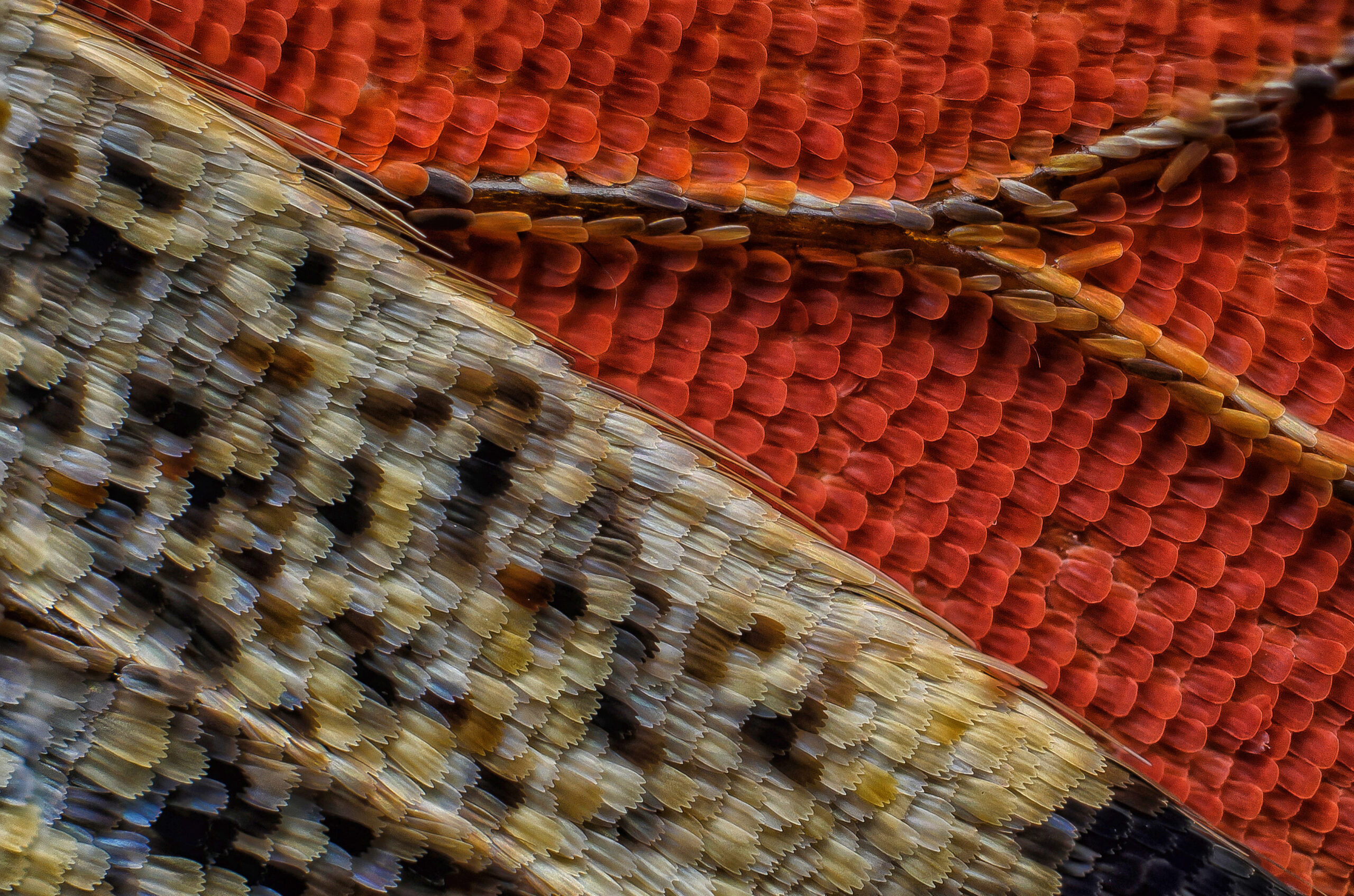
Vein and Scales on a Butterfly Wing: A Tapestry of Beauty
Sébastien Malo, a visionary in the world of microscopy, directs our attention to the microscopic wonders found within the veins and scales of a butterfly wing. Through his exploration, Malo unravels the intricate structures and vibrant colors that adorn these delicate appendages. Under the microscope, butterfly wing veins and scales reveal a mesmerizing mosaic of fine structures and pigments. Each microscopic detail showcases the diversity of wing patterns, the strength of structural components, and the role of pigments in creating the breathtaking visual displays of butterflies. This exploration celebrates the delicate artistry of nature’s designs and the sheer beauty found within the microscopic world of butterfly wings.
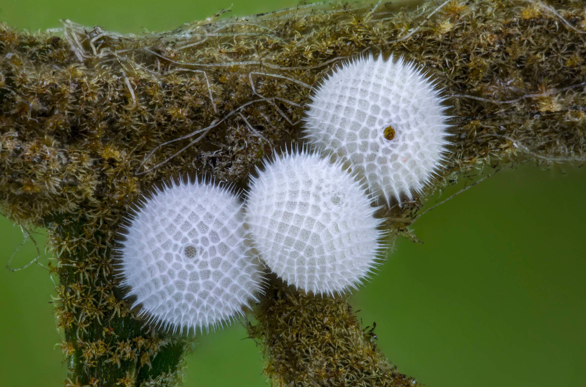
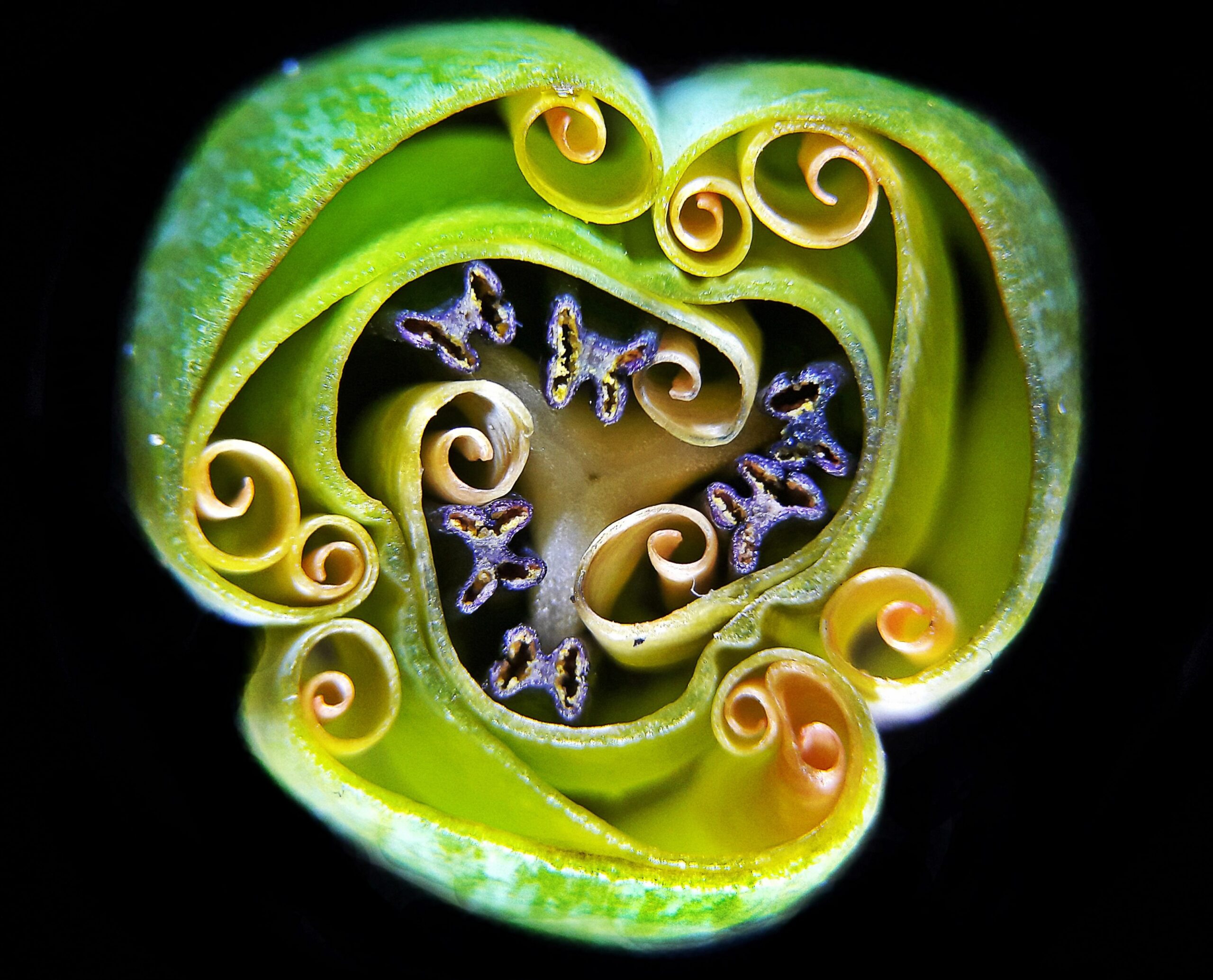
Fern Sori: Unveiling the Reproductive Mysteries
Waldo Nell, a dedicated microscopy enthusiast, directs our attention to the microscopic wonders found within fern sori. Through his lens, Nell unravels the intricate structures and reproductive mysteries that define these ancient plants. Under the microscope, fern sori reveal a tapestry of spore-bearing structures, delicate membranes, and intricate patterns. Each microscopic detail showcases the unique mechanisms employed by ferns to propagate and ensure their survival. This exploration celebrates the marvels of plant reproduction and the enduring legacy of these ancient organisms.
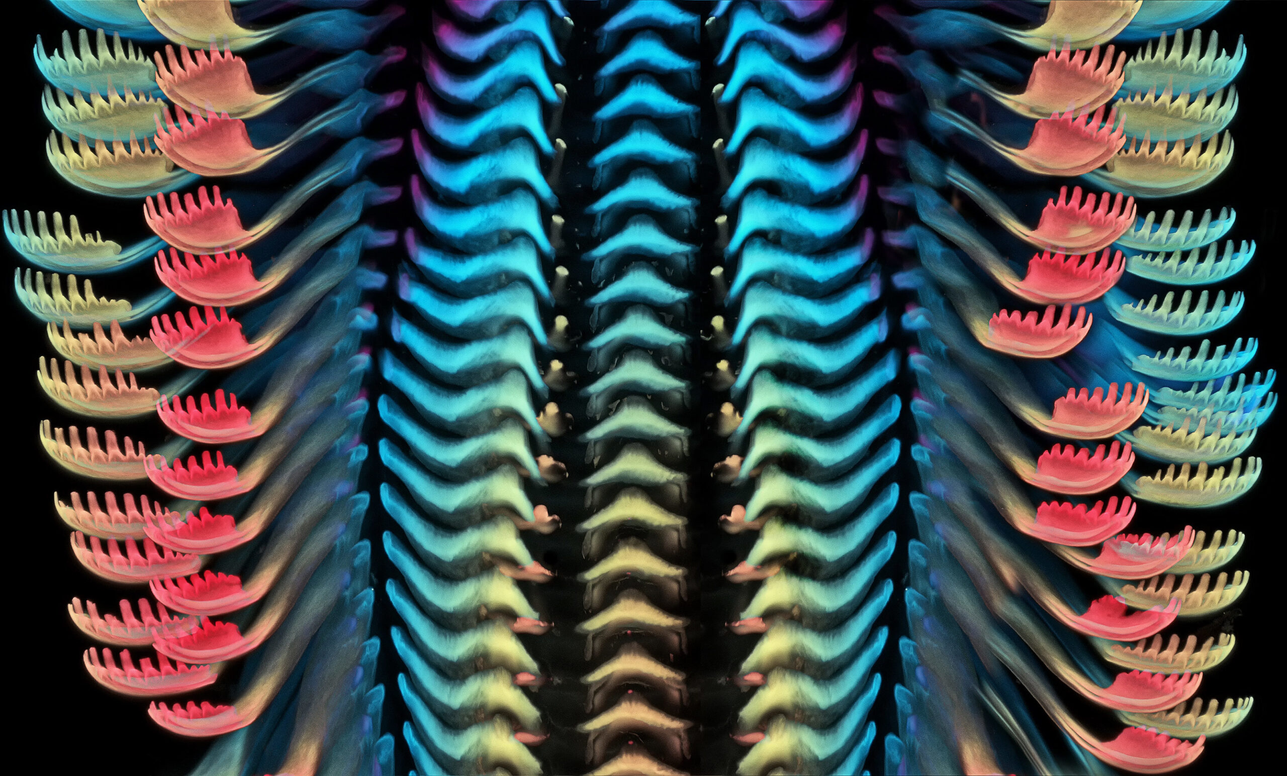
Housefly Eye: A Kaleidoscope of Vision
Dr. Razvan Cornel Constantin, a visionary researcher, focuses his lens on the microscopic wonders found within the compound eye of a housefly. Through his exploration, Constantin unveils the intricate structures and unique visual adaptations that enable these insects to navigate the world. Under the microscope, the housefly eye showcases a mosaic of individual ommatidia, each housing a lens and light-sensitive cells. Each microscopic detail highlights the multifaceted vision and impressive motion detection capabilities of these remarkable insects. This exploration sheds light on the wonders of insect sensory systems and the diverse ways in which creatures perceive their surroundings.
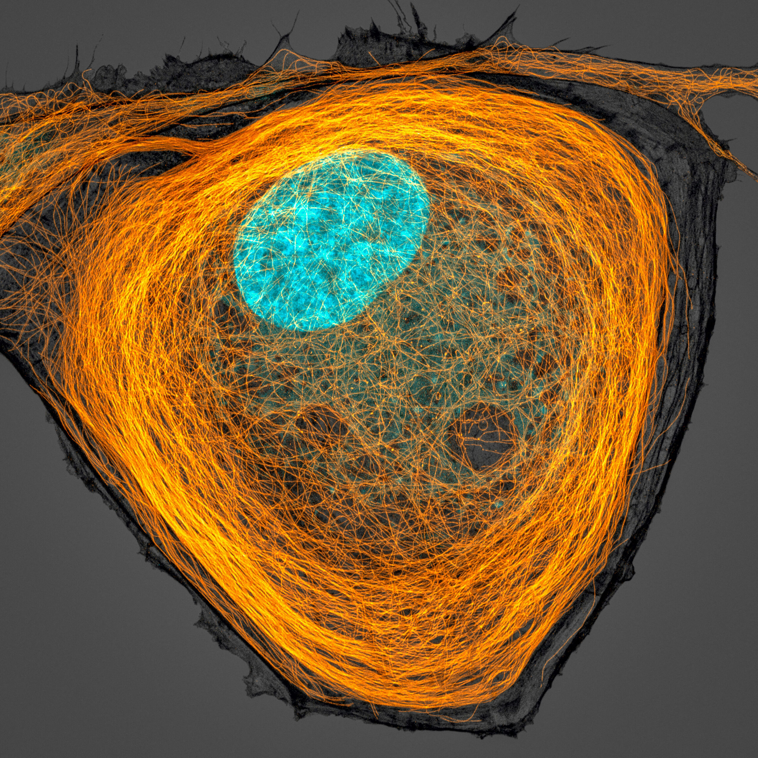
Human Heart Cells: The Rhythm of Life
Hui Lin and Dr. Kim McBride, pioneering researchers in the field of microscopy, invite us to explore the microscopic wonders found within human heart cells. Through their lens, Lin and McBride unravel the intricate structures and coordinated movements that enable the human heart to beat. Under the microscope, human heart cells reveal a network of branching fibers, intercalated discs, and pulsating contractions. Each microscopic detail showcases the remarkable synchronization and coordinated action of these cells, ensuring the continuous flow of blood and the rhythm of life itself. This exploration celebrates the marvels of human physiology and the intricate machinery that keeps our hearts beating.
Flower Crab Spider: Ambush in Living Color
Jorge Perez Carsi, a talented microscopy artist, focuses his lens on the microscopic wonders found within a flower crab spider. Through his exploration, Carsi unveils the intricate structures and vibrant colors that camouflage and aid in the hunting strategies of these cunning arachnids. Under the microscope, the flower crab spider reveals delicate hairs, sharp fangs, and intricate eyes. Each microscopic detail showcases the adaptations that allow these spiders to blend seamlessly with their surroundings and ambush unsuspecting prey. This exploration sheds light on the fascinating world of predator-prey interactions and the remarkable diversity of arachnid adaptations.
Human Lung Cell: The Breath of Life
Joe McKellar, a passionate researcher, directs our attention to the microscopic wonders found within human lung cells. Through his lens, McKellar unravels the intricate structures and delicate membranes that enable our lungs to perform the vital function of oxygen exchange. Under the microscope, human lung cells reveal a complex network of branching airways, thin alveolar sacs, and intricate capillary beds. Each microscopic detail showcases the efficient exchange of oxygen and carbon dioxide that takes place within these specialized cells, allowing us to breathe and sustain life. This exploration highlights the marvels of respiratory physiology and the delicate balance required for optimal lung function.
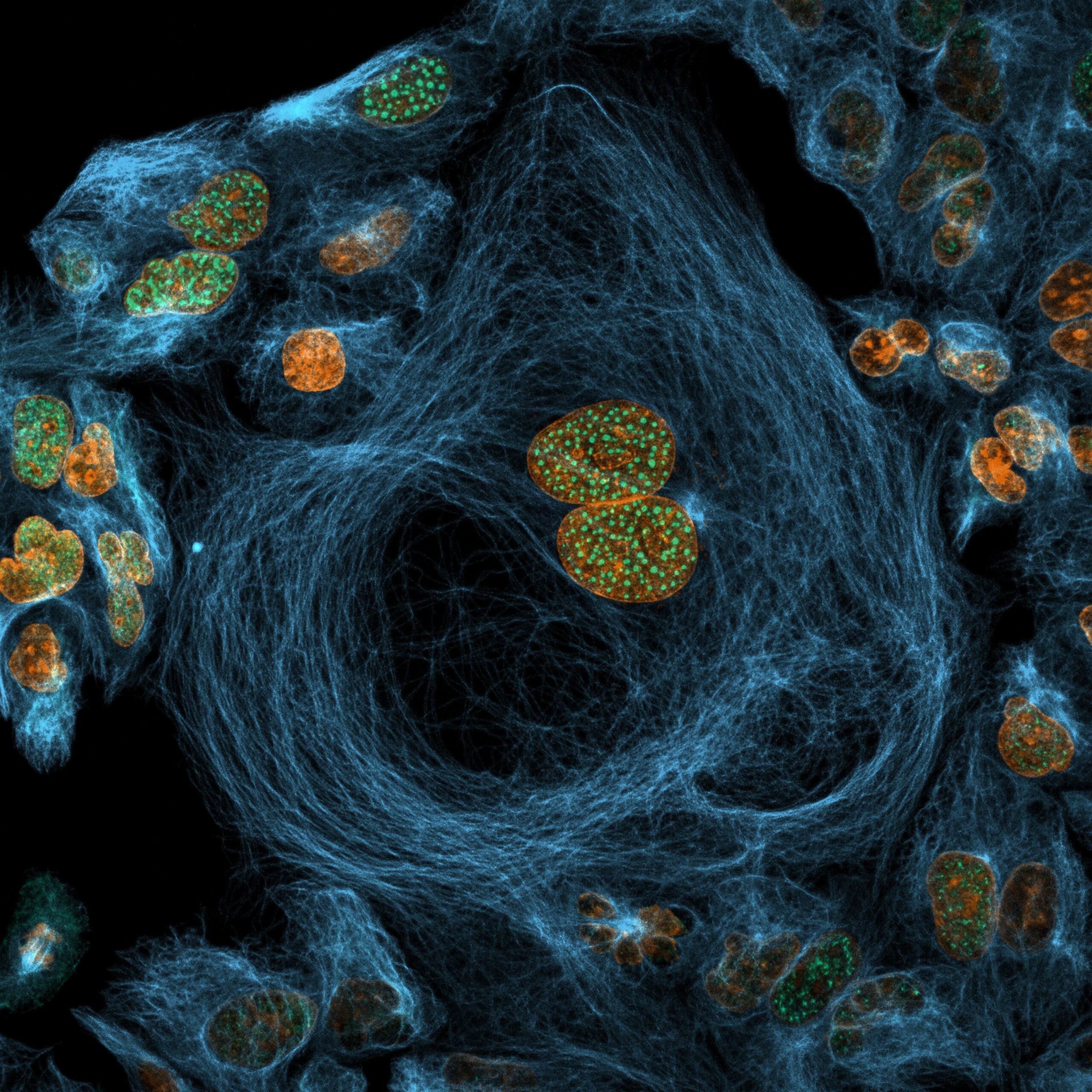
Epithelial Cells: The Building Blocks of Tissues
Caleb Dawson, a dedicated microscopy enthusiast, directs our attention to the microscopic wonders found within epithelial cells. Through his lens, Dawson unravels the intricate structures and essential functions that these cells perform in the human body. Under the microscope, epithelial cells reveal a tightly-packed arrangement, forming the protective lining of organs, glands, and body cavities. Each microscopic detail showcases the specialized shapes and intercellular connections that contribute to the formation of tissues and the maintenance of bodily functions. This exploration highlights the remarkable complexity and importance of epithelial cells in our overall health and well-being.
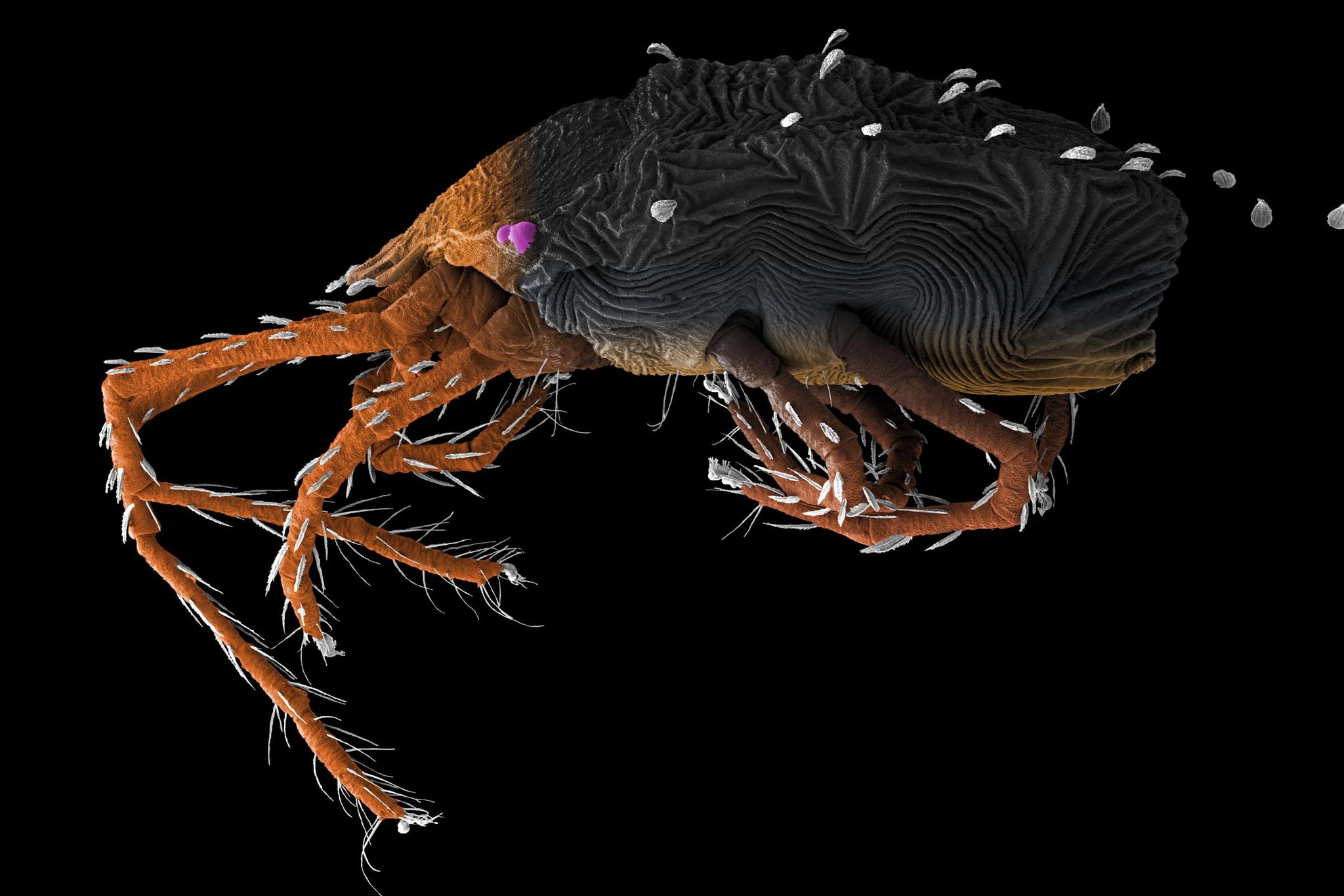
Buckeye Dragon Mite: A Microscopic Marvel
The Buckeye Dragon Mite, captured through the lens of a talented microscopy artist, invites us into the world of these tiny arthropods. Through this exploration, we uncover the microscopic wonders and unique adaptations of these minuscule creatures. Under the microscope, the Buckeye Dragon Mite reveals its intricately segmented body, long appendages, and intricate mouthparts. Each microscopic detail showcases the remarkable adaptations that enable these mites to thrive in their specific ecological niches. This exploration sheds light on the incredible diversity and resilience of nature’s smallest inhabitants.
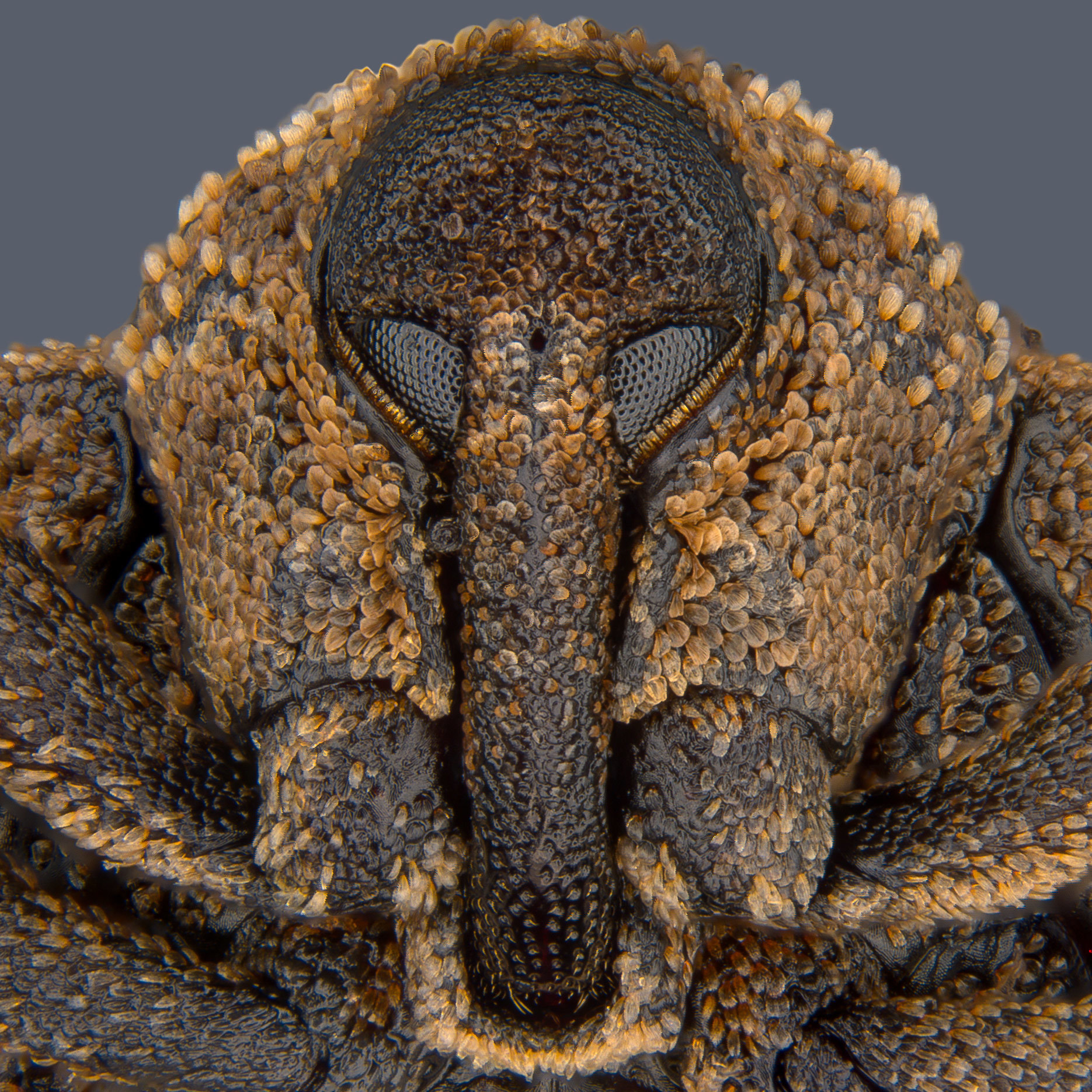
Portrait of Mango Seed Weevil: A Microcosm of Nature’s Intricacy
Pia Scanlon, a visionary in the world of microscopy, presents us with a stunning portrait of the Mango Seed Weevil. Through her lens, Scanlon unveils the microscopic intricacies and captivating features of this fascinating insect. Under the microscope, the Mango Seed Weevil reveals its finely sculpted body, intricate mouthparts, and delicate sensory appendages. Each microscopic detail showcases the adaptations that allow these weevils to feed on mango seeds and complete their life cycle. This exploration celebrates the intricate interplay between insects and plants, highlighting the hidden beauty found within nature’s evolutionary dance.
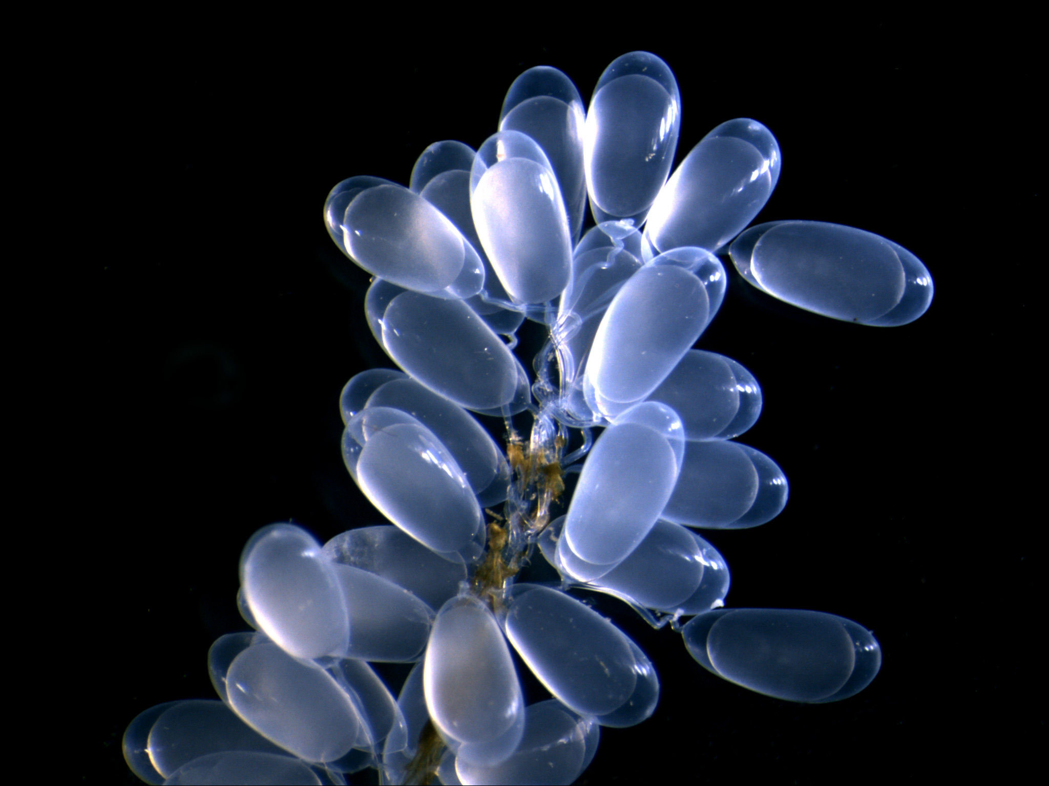
Midget Octopus Eggs: The Miracle of Life Unveiled
Erica Donlon, a passionate microscopy enthusiast, directs our attention to the microscopic wonders found within the eggs of a midget octopus. Through her lens, Donlon reveals the enchanting world of early embryonic development and the miracles that unfold within these tiny spheres. Under the microscope, midget octopus eggs showcase delicate structures, intricate cell divisions, and the development of tiny tentacles. Each microscopic detail captures the essence of new life, highlighting the incredible journey from a single cell to a fully formed organism. This exploration reminds us of the wonder and beauty of the natural world, even at its tiniest scales.
Microtubules: The Cytoskeletal Architects
Jason Kirk, a visionary researcher, focuses his lens on the microscopic wonders found within microtubules. Through his exploration, Kirk unravels the intricate structures and vital roles that these cylindrical protein filaments play within cells. Under the microscope, microtubules reveal their tubular structure and dynamic arrangements, forming highways for molecular transport and providing structural support within cells. Each microscopic detail showcases the architectural prowess of these cellular components and their involvement in crucial cellular processes, including cell division and intracellular transport. This exploration sheds light on the fascinating world of cellular biology and the remarkable organization that underlies life itself.
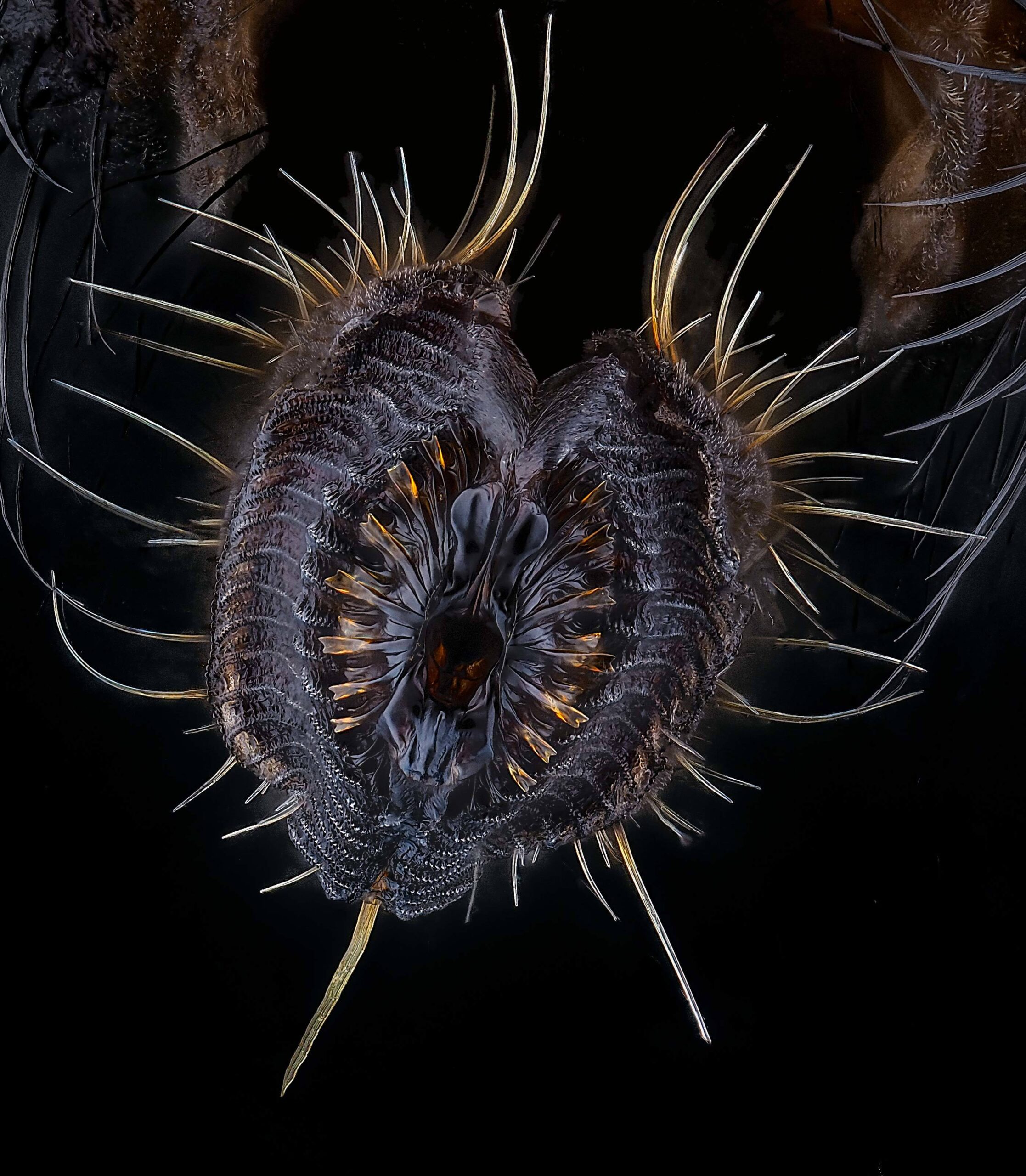
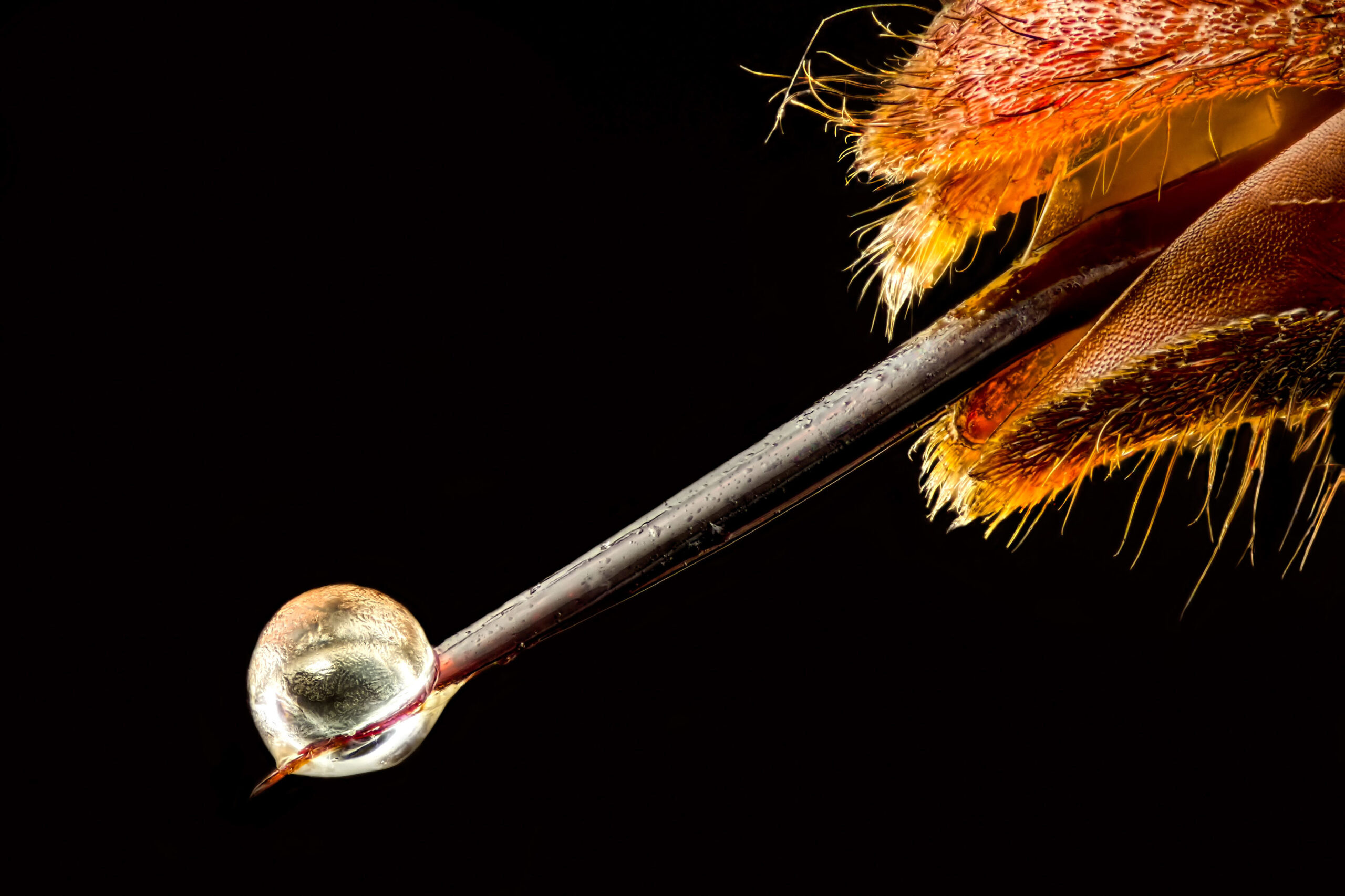
Stinger of a Small Paper Wasp: A Microscopic Weapon
Pablo Piedra, a skilled microscopy artist, invites us to explore the microscopic wonders found within the stinger of a small paper wasp. Through his lens, Piedra unravels the intricate structures and formidable weaponry possessed by these tiny insects. Under the microscope, the wasp’s stinger reveals a sharp, barbed structure designed for defense and subduing prey. Each microscopic detail showcases the adaptations that allow these wasps to effectively inject venom and protect their colonies. This exploration highlights the remarkable defensive mechanisms found within the natural world, even at its smallest scale.
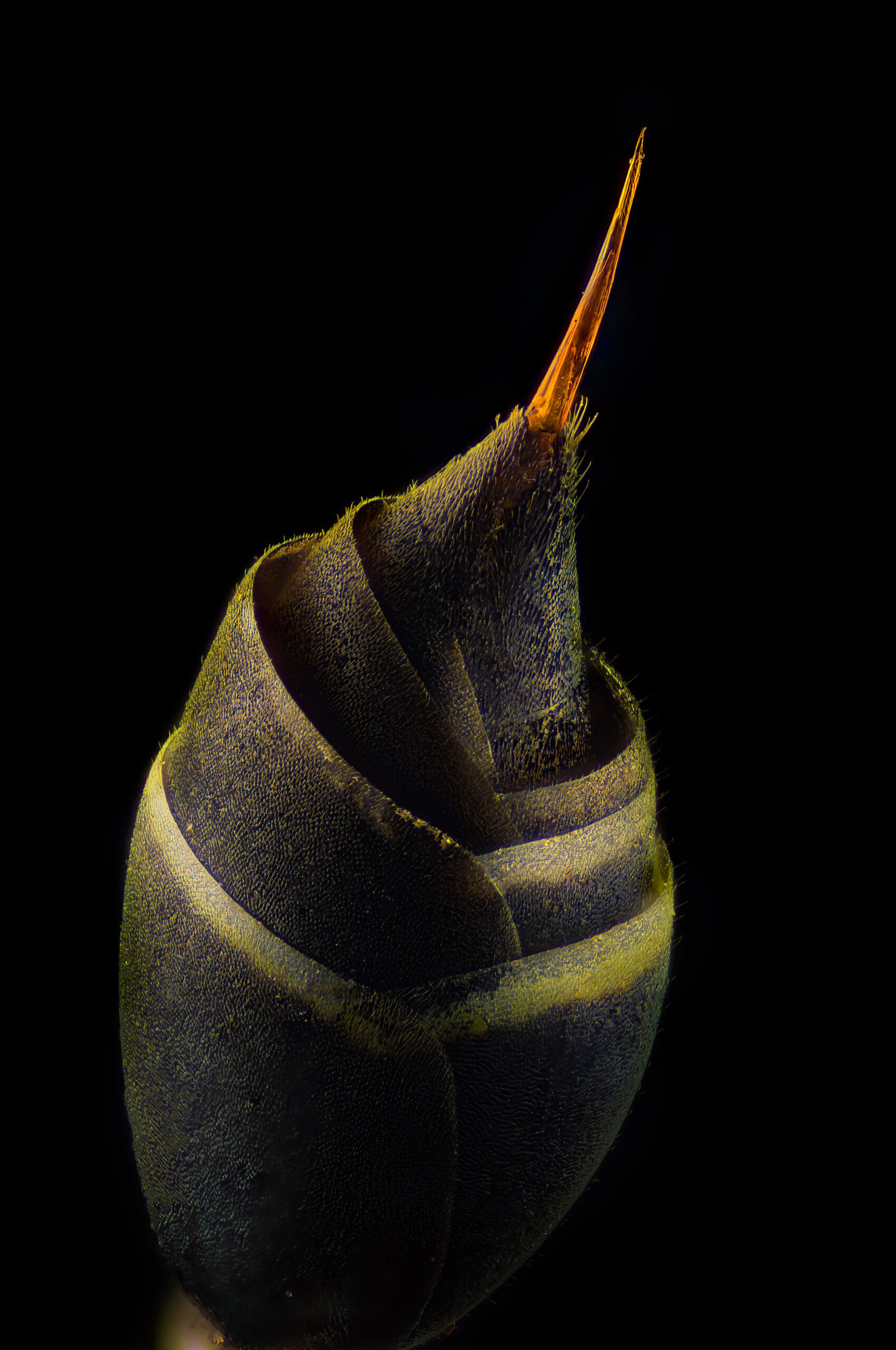
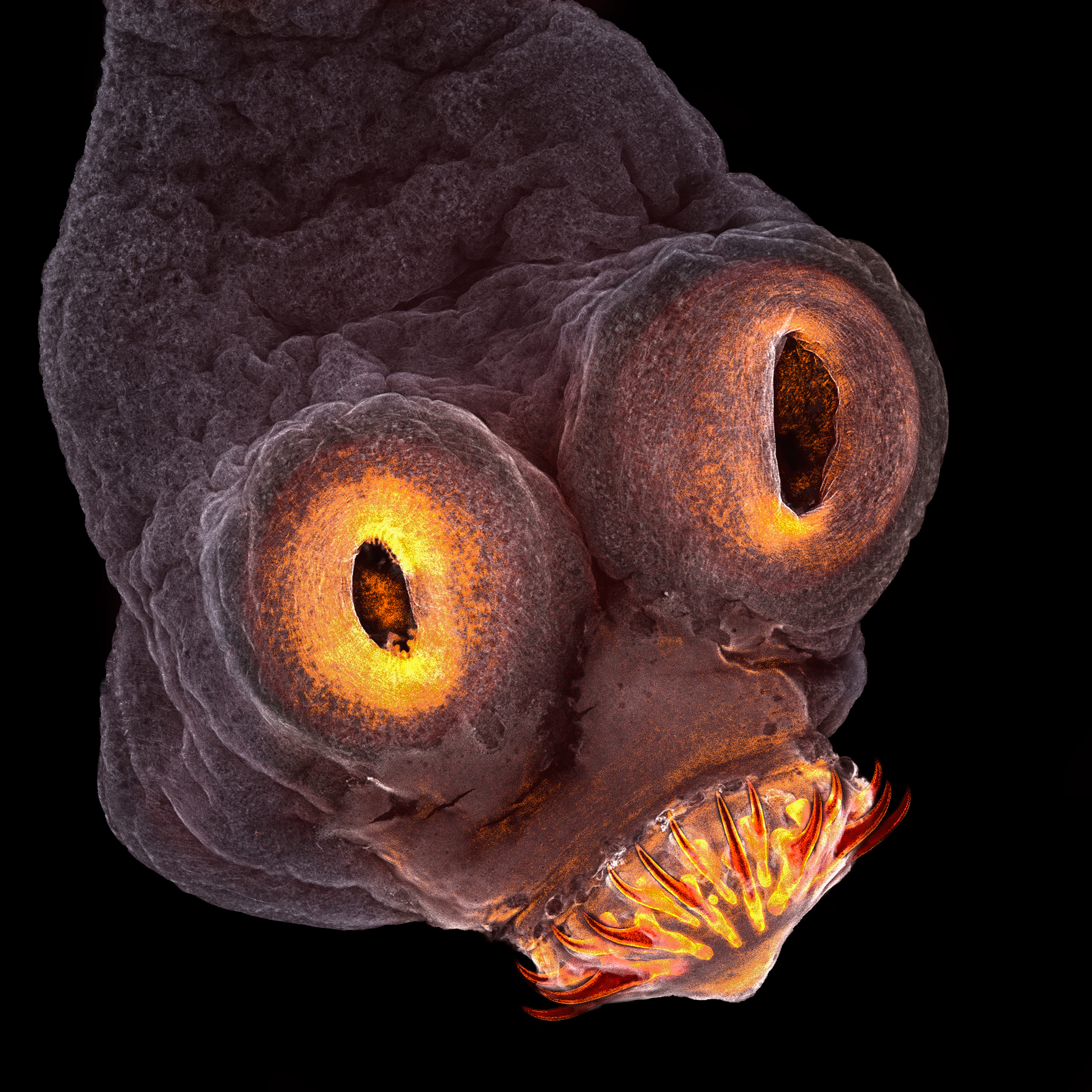
Hedgehog Flea: A Tiny Parasitic Marvel
Dr. Igor Siwanowicz, a renowned researcher, focuses his lens on the microscopic wonders found within a hedgehog flea.Through his exploration, Siwanowicz unveils the intricate structures and remarkable adaptations of these minuscule parasites. Under the microscope, the hedgehog flea showcases its specialized mouthparts, sharp claws, and intricate exoskeleton. Each microscopic detail highlights the adaptations that allow these fleas to cling to their hosts and feed on blood. This exploration sheds light on the intricate interactions between parasites and their hosts, offering a glimpse into the hidden world of microscopic life.
Espresso Coffee Crystals: The Artistry of Aroma
Vin Kitayama and Sanae Kitayama, passionate microscopy enthusiasts, direct our attention to the microscopic wonders found within espresso coffee crystals. Through their lens, they unveil the mesmerizing structures and intricate patterns that give rise to the beloved aroma and flavor of our favorite caffeinated beverage. Under the microscope, espresso coffee crystals reveal their unique shapes and intricate arrangements, showcasing the rich complexity and interplay of compounds that contribute to the distinctive taste and aroma of coffee. Each microscopic detail celebrates the artistry and science behind the perfect cup of espresso, highlighting the beauty found within our morning rituals.
European Pear Rust Fungus: Unveiling a Tiny Pathogen
Dr. Csaba László Pintér, a dedicated microscopy researcher, focuses his lens on the microscopic wonders found within the European pear rust fungus. Through his exploration, Pintér unravels the intricate structures and life cycle of this plant pathogen. Under the microscope, the European pear rust fungus reveals its spores, hyphae, and reproductive structures, showcasing its role in causing devastating infections on pear trees. Each microscopic detail sheds light on the mechanisms of fungal infection and the ongoing battle between plants and pathogens. This exploration highlights the importance of understanding plant diseases and developing effective strategies for crop protection.

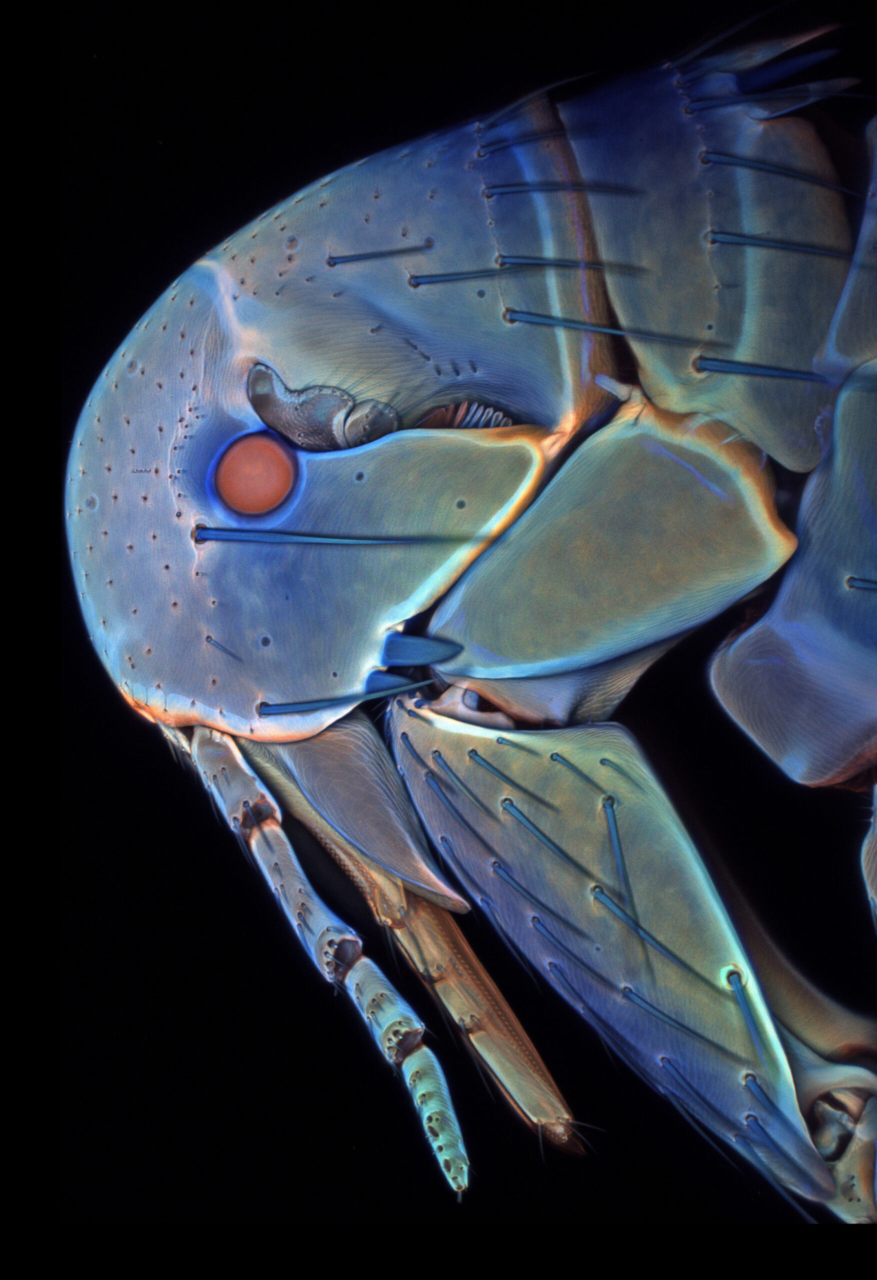
Clover Mite: Nature’s Tiny Wanderer
The Clover Mite, captured through the lens of the USDA’s microscopy experts, invites us to marvel at the microscopic wonders of this tiny arachnid. Through this exploration, we uncover the intricate structures and remarkable adaptability of these wandering creatures. Under the microscope, the Clover Mite reveals its delicate limbs, segmented body, and intricate mouthparts. Each microscopic detail showcases the unique adaptations that enable these mites to navigate diverse environments and feed on plant material. This exploration reminds us of the incredible diversity of life forms that exist even in the tiniest corners of our world.
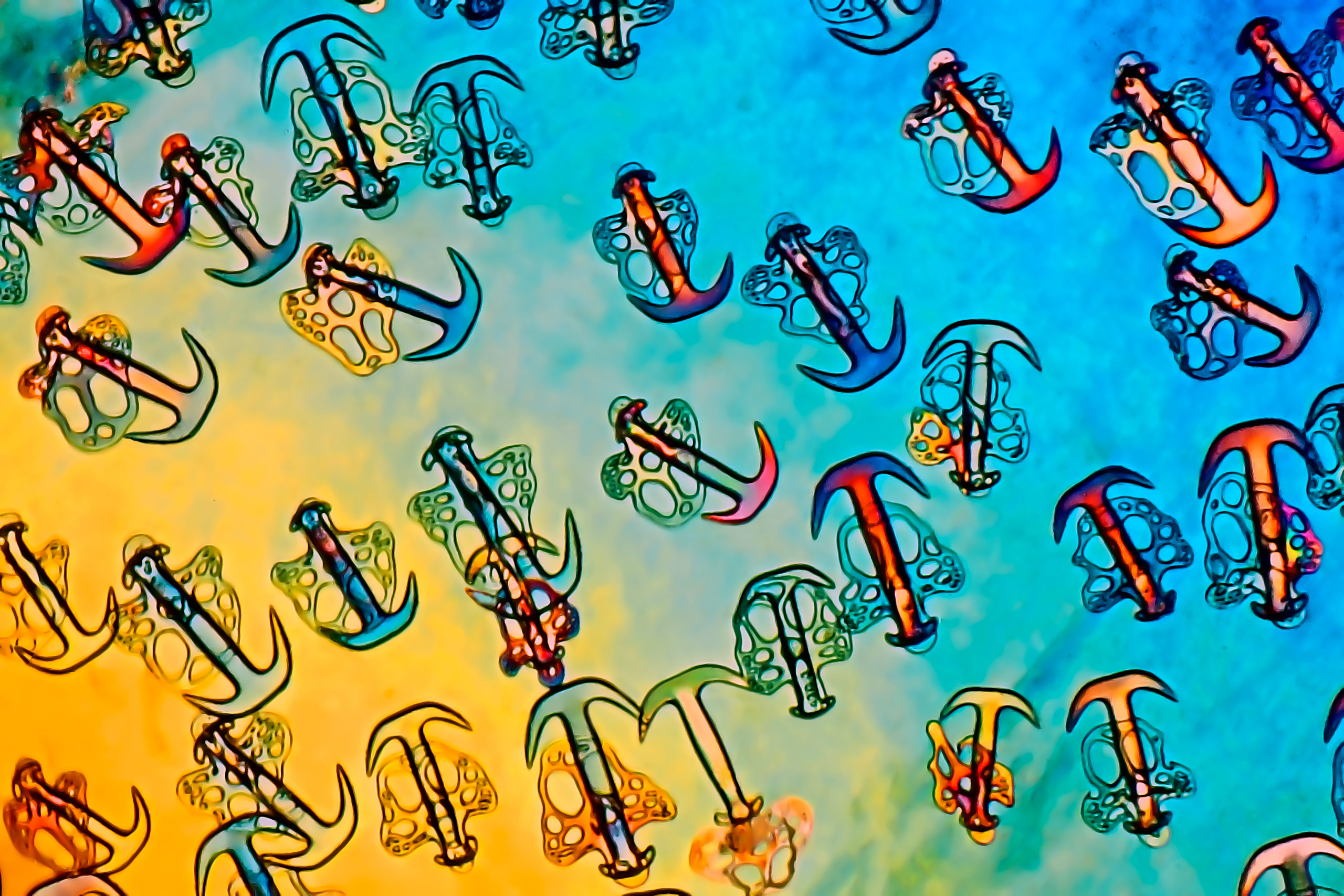
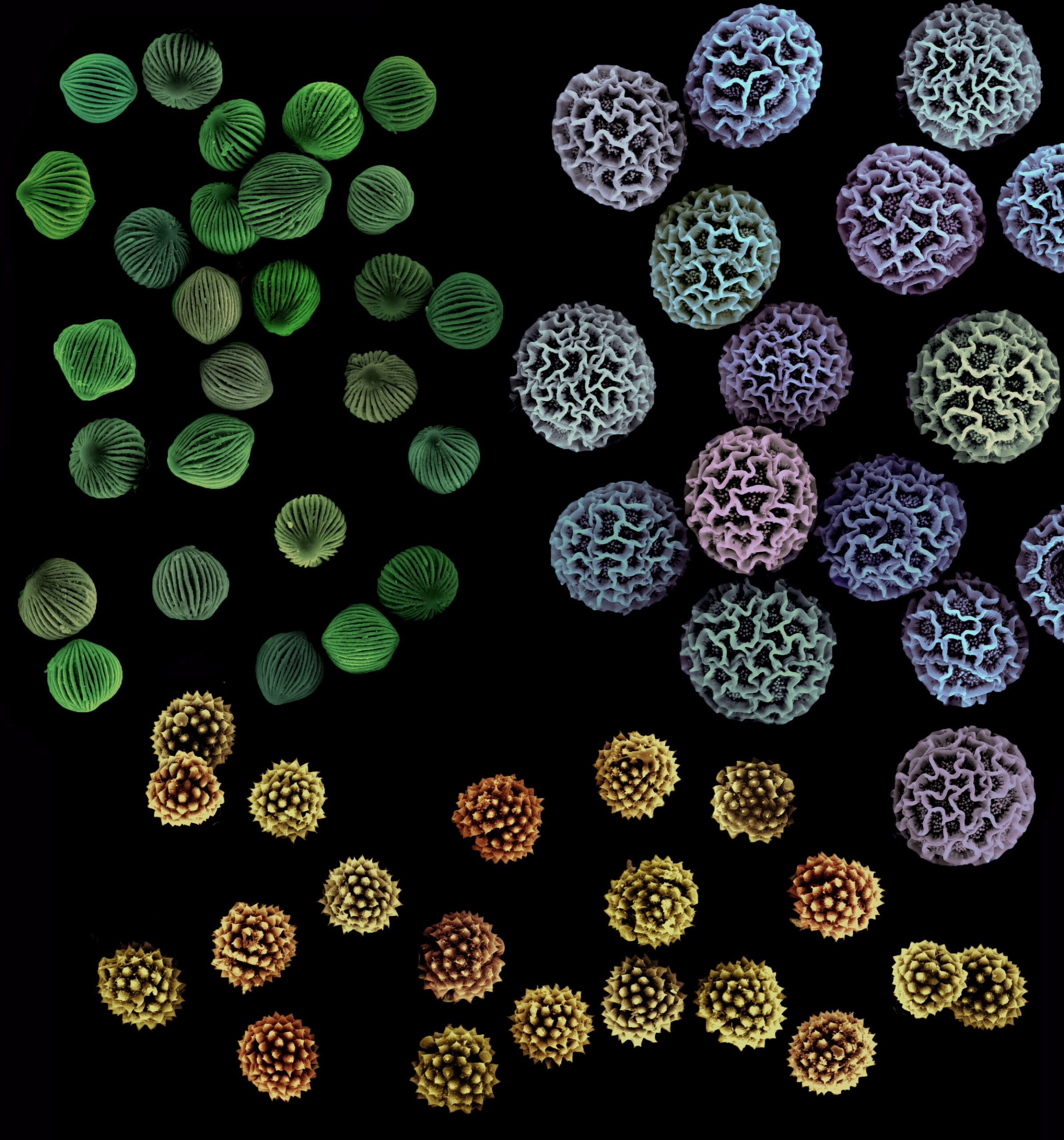
Pollen Grains: Nature’s Tiny Messengers
Within the intricate world of flowers, pollen grains play a crucial role in reproduction. These microscopic marvels, carried by wind or pollinators, hold the potential for new life and vibrant blooms. Explore the diverse shapes, sizes, and textures of pollen grains, and uncover the captivating story they tell about our natural ecosystems.
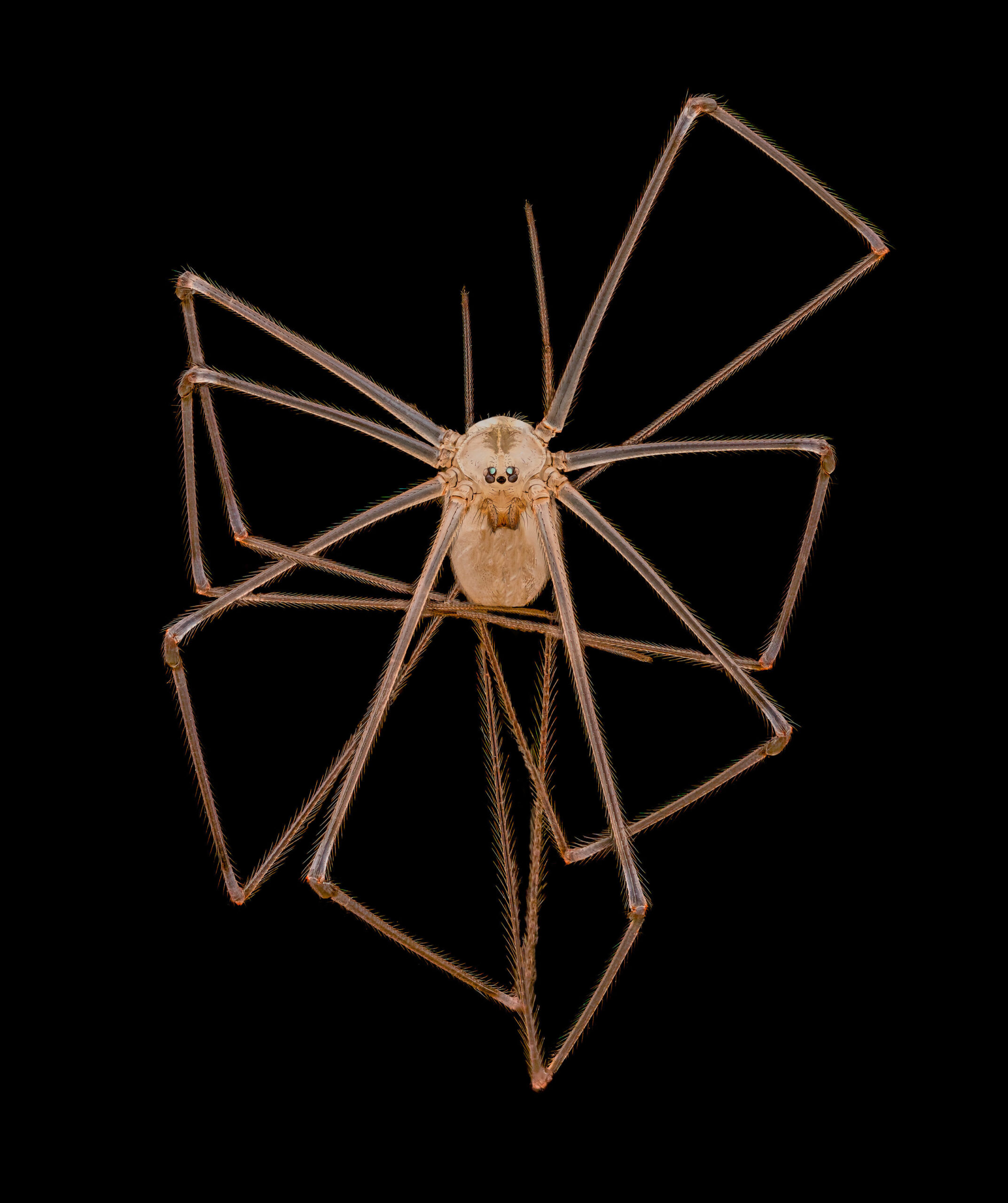
Cellar Daddy Long-legs Spider: A Hidden Resident
Dr. Andrew Posselt delves into the hidden world of the cellar daddy long-legs spider. Through his lens, we witness the intricate anatomy and behavior of these elusive arachnids. Unravel the secrets of their long legs, delicate webs, and unique adaptations that allow them to thrive in the depths of our homes.
Rose Bud Mite: The Stealthy Intruder
The rose bud mite, captured through meticulous observation, reveals its cunning ways. Teresa Zgoda, a skilled microscopy artist, highlights the microscopic intricacies of this tiny pest that infiltrates rose buds, disrupting their growth. Dive into the world of these unseen invaders and discover the battle between nature and pest.
Tapeworm: The Elusive Parasite
Teresa Zgoda focuses her lens on the hidden world of the tapeworm. Under the microscope, this parasitic organism unveils its complex anatomy and life cycle. Discover the adaptations that enable tapeworms to survive within their hosts, and gain insights into the ongoing struggle between parasites and their hosts.
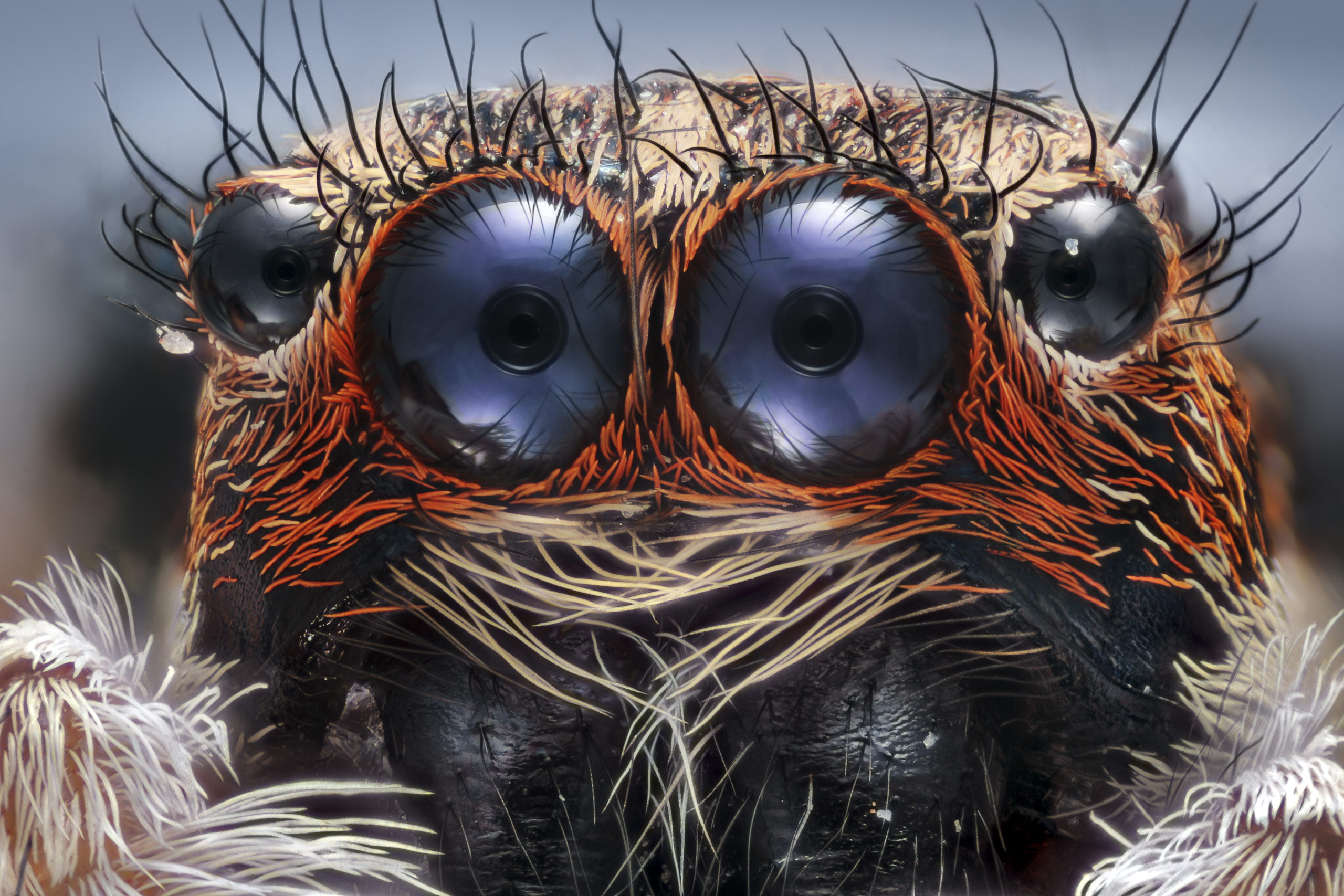
Jumping Spider: Acrobats of the Microcosm
Dr. Andrew Posselt captures the fascinating world of jumping spiders. With their impressive vision, agility, and vibrant colors, these small arachnids defy expectations. Witness their acrobatic leaps, intricate courtship dances, and unique hunting techniques through the lens of a microscope. Prepare to be amazed by the beauty and versatility of these charismatic arachnids.